This article has been reviewed according to Science X's editorial process and policies. Editors have highlighted the following attributes while ensuring the content's credibility:
fact-checked
trusted source
proofread
Advanced 3D imaging techniques boost understanding of root systems for food security

The increasing frequency of extreme weather events and global population growth present challenges to food security, emphasizing the need for resilient and high-yielding crops. Central to this challenge is understanding Root System Architecture (RSA), influenced by soil conditions.
Ongoing research is dedicated to overcoming hurdles in root phenotyping, with a particular focus on three-dimensional (3D) imaging of soil-grown roots utilizing magnetic resonance imaging (MRI).
Despite notable progress, challenges persist, including issues like low contrast in MRI images and the complexity of segmentation, which hinder accurate RSA analysis. Recent advancements in deep learning, such as the 3D U-Net, show potential in enhancing image segmentation and analysis, crucial for effective RSA study in varying soil conditions.
In July 2023, Plant Phenomics published a research article titled "3D U-Net Segmentation Improves Root System Reconstruction from 3D MRI Images in Automated and Manual Virtual Reality Work Flows."
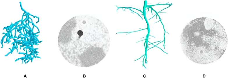
This study introduced an innovative two-step automated workflow for the reconstruction of MRI-based root systems, aiming to overcome previous challenges in the field. The first step involved applying a 3D U-Net, developed by Zhao and a research team, to enhance contrast-to-noise ratio (CNR) and resolution in MRI images through super-resolution segmentation of roots and soil.
Despite minor gaps and noise, this segmentation improved root visibility. The second step utilized Horn et al.'s automated root reconstruction algorithm, designed for imperfect data, to trace roots from these segmented images.
The efficacy of this workflow was assessed by comparing three reconstruction methods: manual expert reconstructions of raw MRI images (M), manual reconstructions on segmented images from the 3D U-Net (M+), and fully automated reconstructions (A).
These methods were evaluated using MRI scans of lupine plants grown in two different soil substrates, focusing on the visual comparison of reconstructed root systems and the calculation of characteristic root measures. The research aims to determine if 3D U-Net segmentation can recover more root length in manual reconstructions and if the automated workflow can produce tracings of comparable quality.
Results showed that M+ reconstructions generally included more roots and slightly longer root lengths compared to M, particularly in first-order roots. The similarity between roots identified in both manual methods was high, indicating that working on segmented images does not significantly impact human decision-making. However, the mean root radius was typically larger in M+ reconstructions.
The A method showed lower total root length compared to M and M+ and displayed more frequent directional changes in root trajectories, suggesting difficulties in bridging large gaps in U-Net segmentation. A sometimes differed in root connectivity and topological accuracy. Quantitatively, the root measures supported these observations, with variations in root length recovery rates and root radii among the methods.
The U-Net segmentation notably improved reconstruction rate and root recovery in low-CNR data. Automated reconstructions provided similar root metrics to manual methods, especially in high-CNR scenarios, although they still face challenges in topological decision-making and gap closing.
In summary, the study demonstrates that 3D U-Net segmentation significantly enhances manual reconstruction workflows, particularly in low-CNR conditions, by improving root visibility and reducing reconstruction time. While the automated reconstruction method shows promise, it requires further refinement in handling topological complexities and gap closures.
This research contributes to advancing automated root system reconstruction methodologies, particularly in challenging MRI environments.
More information: Tobias Selzner et al, 3D U-Net Segmentation Improves Root System Reconstruction from 3D MRI Images in Automated and Manual Virtual Reality Work Flows, Plant Phenomics (2023). DOI: 10.34133/plantphenomics.0076
Provided by NanJing Agricultural University


