This article has been reviewed according to Science X's editorial process and policies. Editors have highlighted the following attributes while ensuring the content's credibility:
fact-checked
peer-reviewed publication
trusted source
proofread
Whispering gallery microprobe opens up new opportunities for optical spectroscopy

Light–matter interaction is one of the most basic ways people observe the physical world. While reflection and refraction of light reveal the morphology of matter, inelastic scattering of light, like Raman scattering, encodes the molecular fingerprint of chemical bonds into the energy shift of photons. However, the possibility of such interactions is vanishingly small.
Researchers have been developing specific mechanisms and structures to enhance Raman signals over the past decades. Surface-enhanced Raman spectroscopy, or SERS, represents one of the most promising platforms of such efforts, where metallic nanostructures are introduced to significantly enhance the electromagnetic field near the sample surface and also tailor targets' density of energy states in favor of Raman scattering. On the other hand, whispering-gallery-mode (WGM) microresonators have emerged as frontrunners to enhance light-matter interactions in applications ranging from sensing, spectroscopy, imaging, etc.
These sub-millimeter resonators could work as a reservoir of light, accumulating light and realizing a temporally enhanced optical field. The combination of the two platforms is undoubtedly promising and intriguing, providing boosted light-matter interaction both spatially and temporally. However, a systematic investigation and proof-of-concept demonstration was needed.
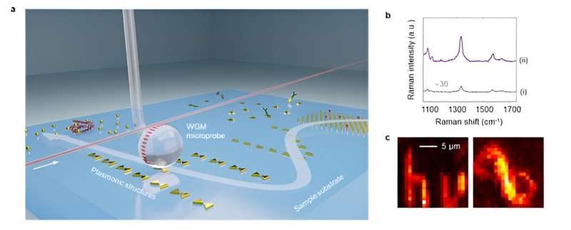
In a recent paper published in Light: Science & Applications, a team of researchers led by Professor Lan Yang from Washington University in St. Louis presented a novel Raman enhancing platform where a WGM microprobe is mated with nano-plasmonic SERS structures. Nano-plasmonic hotspots are coupled with WGMs via a phase-matched cavity-antenna coupling mechanism to maximize the spontaneous Raman scattering from various chemical and biochemical samples.
Two-orders-of-magnitude enhancement was reported on top of the stand-along nano-plasmonic hotspots conventionally excited by a focused free-space light beam. The group also showed the compatibility of the microprobe to different types of SERS substrate, including lithographically defined nano-plasmonic hotspot array and commercial SERS substrate paper, demonstrating the versatility of the novel platform.
More interestingly, by mounting the probe onto a translation stage, a two-dimensional hyperspectral Raman imaging with signal enhancement was realized with only sub-milliwatt continuous-wave pump power. The reported results opened the door to explore inelastic light-matter interaction enhancement with WGM-plasmonic hybrid resonance, and also presented a versatile tool for Raman-based material analysis and chemical imaging.
"This paper is a summary of our group's long journey exploring the combination of WGM microresonators and nanoplasmonic SERS structures. We found that instead of simply serving as an optical pump excitation interface, WGMs form a hybrid mode with nano-plasmonic hotspots. In addition, due to the guided-wave nature of WGMs, we observed manifestation of the phase-matching condition, which is both evidence of the hybrid resonance and also a significant contributor to the Raman signal enhancement that could be leveraged," the authors explained.
"In some ways, the WGM microprobe platform works similarly to an atomic force microscopy (AFM) tip. We also tried to demonstrate and evaluate the potential of this platform from an apparatus development point of view. This motivates our study of enhancing conventional SERS and 2D scanned Raman imaging using the microprobe configuration. With all the results presented, we are confident to say that this new technique could unlock many opportunities for a better and potentially more compact SERS test platform," the team says.
"Probably the over-simplified way to understand our work is: we developed a versatile and scannable contact lens to SERS samples," concluded the researchers.
More information: Wenbo Mao et al, A whispering-gallery scanning microprobe for Raman spectroscopy and imaging, Light: Science & Applications (2023). DOI: 10.1038/s41377-023-01276-2
Journal information: Light: Science & Applications
Provided by Chinese Academy of Sciences


