This article has been reviewed according to Science X's editorial process and policies. Editors have highlighted the following attributes while ensuring the content's credibility:
fact-checked
peer-reviewed publication
trusted source
proofread
Researchers zoom in on new ways to view biomolecules in pathogens

A new set of protocols will allow researchers to expand the way they look at pathogens.
The principle of physically expanding biological study samples for imaging is known as expansion microscopy. The technique uses a hydrogel to homogenously expand cells and decrowd biomolecules, providing researchers with a way to visualize fine details using standardized microscopes instead of expensive, specialized tools.
Led by Eberly Family Career Development Associate Professor of Biological Sciences Leon Zhao, the Zhao Biophotonics Lab at Carnegie Mellon University is a leader in advancing expansion microscopy for biomedical applications.
MicroMagnify, the latest protocol published by the Zhao Lab, takes their work a step forward. The protocol expands complex microbial cells and infected tissues without distortion, allowing for enhanced high-plex fluorescence imaging.
Super-resolution optical imaging tools are crucial in microbiology to understand the complex structures and behavior of microorganisms such as bacteria, fungi and viruses.
"Visualizing infected tissues and pathogens has always been challenging due to the minuscule size of bacterial cells, often 1 micron or less," Zhao explained. "The development of a cost-effective, nanoscale imaging technique to visualize pathogens within host tissue is crucial for both research and clinical applications."
Traditional expansion methods struggle to break down the thick, rigid cell walls of bacterial pathogens like staphylococcus aureus and the similar envelopes of fungal pathogens that protect cell shapes.
However, Carnegie Mellon doctoral student Zhangyu "Sharey" Cheng discovered that a mix of heat denaturation and enzyme cocktails effectively breaks down both types of cellular walls and expands microbial cells and infected tissues without distortion. This process also preserves diverse biomolecules and allows post-expansion staining for imaging. Cheng is first author on a paper outlining the process in Advanced Science.
Researchers often will use fluorescent proteins to track molecules and cells during imaging, but current protocols limit instrumentation to at most five targets in one sitting. With MicroMagnify, Zhao said that they demonstrated how one sample could be used to image 10–12 targets within a pathogen-infected mammalian cell.
"MicroMagnify preserves biomolecules and proteins in the gel, enabling a cycle of imaging, labeling and washing with different reagents each round. Theoretically, this allows the study of hundreds of targets," Zhao added.
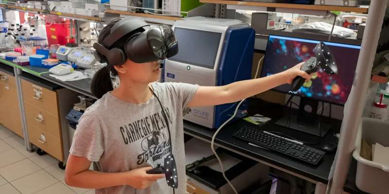
To better understand the complex three-dimensional images generated by MicroMagnify, the Zhao Lab and Benaroya Research Institute (BRI) at Virginia Mason Franciscan Health collaborated on ExMicroVR, an immersive virtual reality environment. With a low-cost VR headset, GPU-accelerated computer and internet access, scientists can meet virtually to explore biological data.
Caroline Stefani, research assistant member at BRI, helped pioneer the use of virtual reality as a 3D visualization tool for fluorescence images and prepared samples for the Zhao Lab. Stefani and Tom Skillman, BRI's former director of research technology, met Zhao in the Grand Challenges Annual Meeting, organized by the Bill & Melinda Gates Foundation.
"In this space, they can see the cell image and interact with simple avatars of each other," said Skillman, who founded Immersive Science, a VR company. "They can manipulate the cell image, point, talk, locate regions of interest and engage in discussions about the cell's response to an invading pathogen."
Next, Cheng is using MicroMagnify to study the microbiomes in the gut. "Microbiomes are incredibly complex and their interactions with host organs can significantly affect disease progress, such as tumors," Cheng said.
"With MicroMagnify methods, we can resolve the composition and signaling of these microbiomes and explore how their interactions with host organs affect disease progression, such as cancer. This will enable us to profile the microbiomes and their signaling proteins with nanoscale precision and identify specific populations that may contribute to disease progression or, conversely, help prevent it."
More information: Zhangyu Cheng et al, MicroMagnify: A Multiplexed Expansion Microscopy Method for Pathogens and Infected Tissues, Advanced Science (2023). DOI: 10.1002/advs.202302249
Journal information: Advanced Science
Provided by Carnegie Mellon University


