This article has been reviewed according to Science X's editorial process and policies. Editors have highlighted the following attributes while ensuring the content's credibility:
fact-checked
trusted source
proofread
Scientists marry MRI, ultrasound, and optoacoustics for improved medical exams

Physicians and researchers rely on biomedical imaging to examine the structure and function of living tissue. This enables disease diagnostics and experiments that reveal the mechanisms behind pathologies and ways to treat them. The most popular techniques for radiation-free imaging are ultrasound and MRI scans. Optoacoustics, on the other hand, is a promising emerging approach only recently introduced into clinical practice.
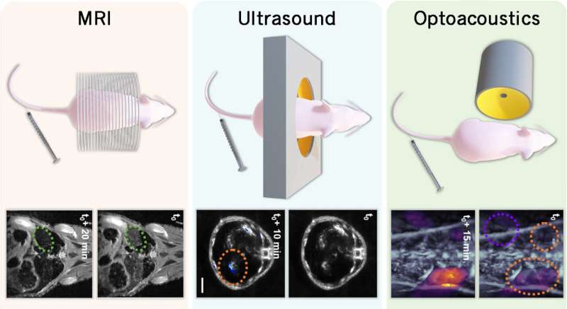
Now, Skoltech researchers and their Swiss and Chinese colleagues have managed to marry these distinct imaging techniques by devising a universal contrast agent—an injectable drug that simultaneously works with all three approaches. The new agent could make diagnostics faster and more accurate, while reducing examination cost, the number of injections, and the dosage necessary.
Besides enabling high-contrast visualization, the team's "loaded microbubbles" could even be used in the future to deliver drugs into the brain of a patient with Parkinson's or a tumor. The findings are reported in Laser & Photonics Reviews.
The researchers used a technology known as layer-by-layer deposition to make microbubbles loaded with indocyanine green dye and magnetite nanoparticles. The dye can absorb light and emit detectable sound waves, which is how optoacoustics works. And the nanoparticles of magnetite, an oxide of iron, enhance contrast during MRI exams. The bubbles themselves serve as a contrast agent for ultrasound studies, and because they are filled with liquid—a nanodroplet of perfluoropentane—rather than gas, increased stability is achieved.
The team carried out experiments on mice and made sure that the microbubbles exhibited contrast in all three modes of medical imaging. Cytotoxicity tests showed the agent is biocompatible.
"The individual contrast agents used in any given imaging technique have their advantages, but by bringing them together we make them complement each other. This translates, among other things, into higher sensitivity and better imaging resolution. And we reduce invasiveness, because where you used to require three separate injections, now you only need one," one of the study's two lead authors, Daniil Nozdriukhin, said.
"Also, with the microbubbles, the circulation times of both the nanoparticles and the dye in the body are way longer, which means there is more time to get a high-quality image. The stability and longevity of the liquid-core bubbles is an added benefit on top of that."

A further tentative application of the new contrast agent is magnetic resonance and optoacoustic imaging of the brain. The problem with visualizing the brain is that the so-called blood-brain barrier only allows a select few molecules from the bloodstream to enter the brain: oxygen, nutrients, hormones, etc.
The barrier shuts out all manner of germs and large molecules, including contrast agents and most drugs. It can be opened by generating gas bubbles inside blood vessels with ultrasound. This, however, harms the surrounding tissue. Fortunately, it is possible to make do with a much lower intensity by using focused ultrasound on microbubbles, and this is where the team's multifunctional contrast agent comes in.
"With one agent bringing together both the microbubbles, sensitive to ultrasound, for opening the blood-brain barrier and the contrast materials for MRI and optoacoustic imaging, a single injection will suffice for a brain exam, and you get the additional benefit of extended circulation into the bargain," lead author of the study, Elizaveta Maksimova said.
"What's more, the liquid-core microbubbles can withstand ultrasound exposure without bursting for much longer than the conventional gas-core microbubbles, holding the barrier open for prolonged periods of time so that the dose of the contrast agent in the injection can be lowered."
"Also, once you have this effective and safe way to open the blood-brain barrier, you can go beyond pure diagnostics and enhance the bubbles by loading them with a drug via the same layer-by-layer deposition approach. Such integration of therapeutic agents and those used for diagnostics is known as theranostics," added the study's principal investigator Professor Dmitry Gorin, who heads the Biophotonics Lab at Skoltech Photonics.
"This approach can be applied for the MRI-guided minimally invasive treatment of glioblastoma [the most aggressive and most common type of cancer that originates in the brain]."
How layer-by-layer deposition works
Bubbles filled with perfluoropentane—a liquid at room temperature—are stabilized with a protein and immersed in a series of water solutions. The particles from each successive solution are deposited as an additional shell on the microbubble, provided that compounds with positively and negatively charged inorganic particles or organic molecules are alternated.
The electrostatic interaction holds the shells together. In the study reported in this story, the deposited layers contained contrast agents for MRI and optoacoustic imaging, but the same procedure can be used with therapeutic agents.
More information: Elizaveta A. Maksimova et al, Multilayer Polymer Shell Perfluoropentane Nanodroplets for Multimodal Ultrasound, Magnetic Resonance, and Optoacoustic Imaging, Laser & Photonics Reviews (2023). DOI: 10.1002/lpor.202300137
Provided by Skolkovo Institute of Science and Technology


