This article has been reviewed according to Science X's editorial process and policies. Editors have highlighted the following attributes while ensuring the content's credibility:
fact-checked
peer-reviewed publication
proofread
Mapping the lamellar structure of human corneas using polarization-resolved-SHG

An essential role of the cornea is to contribute to light focusing on the retina. Its curved shape, and therefore its ability to focus light, must be perfectly stable to ensure a sharp image despite the various shocks and rubs received over the course of a day. The cornea is thus composed of several hundred collagen lamellae stacked one on top of the other with various in-plane orientations.
But the transparency of this tissue, essential for visual acuity, is an obstacle to optical imaging of its structure using conventional optical microscopes. As a result, poor characterization of the lamellar structure of the cornea today limits our understanding of the link between structure and function of this tissue, particularly in terms of its mechanical properties. Another limitation concerns the understanding of certain pathologies linked to a defective structure, such as keratoconus.
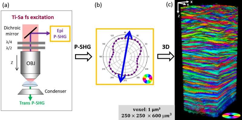
In a new paper published in Light: Science & Applications, a team of scientists, led by Professor Marie-Claire Schanne-Klein from the Laboratory for Optics and Biosciences at Ecole Polytechnique, CNRS, Inserm and Institut Polytechnique de Paris, Palaiseau, France, have used a recent three-dimensional (3D) optical imaging technique: second harmonic generation (SHG) microscopy, which enables specific visualization of collagen without any prior labeling.
The originality of their approach lies in playing on the polarization of light to reveal the direction of the collagen fibrils that compose the lamellae of the cornea. This imaging is performed in reflection, using epi-detection and may be further developed to eventually enable in vivo diagnosis. Another advantage of this configuration is that it provides far more accurate results than transmission.
This is because the coherence length of SHG processes is shorter in the backward direction than in the forward direction, which in practice ensures better spatial resolution. Ten intact human corneas were thus characterized. A careful validation was performed by two self-referenced methods, as SHG polarimetry had never been implemented on tissue of such thickness (around 600 µm).
In particular, the authors have verified that the same structure is obtained by imaging the cornea in two different orientations. This study has thus shown for the first time that, while the lamellae of the human cornea are globally oriented in two perpendicular directions, their main direction changes with depth.
This study paves the way for promising new characterizations of the cornea: mapping the size and distribution of lamellae as a function of depth, but also as a function of position (center versus periphery of the tissue). This information will feed into mechanical modeling of corneal behavior in response to variations in intraocular pressure or healing processes. Finally, the study of pathological tissues will enable to establish the role of corneal structure in certain diseases.
More information: Clothilde Raoux et al, Unveiling the lamellar structure of the human cornea over its full thickness using polarization-resolved SHG microscopy, Light: Science & Applications (2023). DOI: 10.1038/s41377-023-01224-0
Journal information: Light: Science & Applications
Provided by Light Publishing Center, Changchun Institute of Optics, Fine Mechanics And Physics, CAS


