This article has been reviewed according to Science X's editorial process and policies. Editors have highlighted the following attributes while ensuring the content's credibility:
fact-checked
trusted source
proofread
Triple study exploits cellular messengers for diagnostics and drug delivery

In a series of three papers on extracellular vesicles (EVs)—particles that cells use to communicate—Skoltech researchers and their colleagues have presented two methods for extracting EVs from blood samples for diagnosing certain types of cancer and other diseases, as well as a technique for coaxing cells into "taking the medicine" by making drug molecules appear like EVs. The studies were published in the Journal of Extracellular Vesicles, Analysis & Sensing and Colloids and Surfaces B: Biointerfaces.
Cells are pretty sociable, and one of the ways they contact one another is by dispatching particles called extracellular vesicles. EVs are enclosed in a membrane and carry messages in the form of a "soup" of nucleic acids and proteins on the inside, as well as proteins on the surface—the latter are studied by Skoltech photonics researchers from the Biophotonics Lab headed by Professor Dmitry Gorin.
What makes extracellular vesicles interesting is the possibility of using them for medical diagnostics and therapy. Because EVs produced by different cells—e.g., cancer cells—can be distinguished by characteristic surface proteins, they can be used for early diagnostics of their associated diseases, including cancer, infectious diseases and others. As for therapy, EVs are recognized by the cell as messengers and freely admitted inside, so it makes a lot of sense to try and pack drug molecules in them.
Skoltech Senior Research Scientist Vasily Chernyshev and his colleagues from MIPT and Skolkovo-resident startup Prostagnost have developed a way to isolate extracellular vesicles from blood plasma and other biological fluids that yields more EVs with greater purity than the two techniques commonly used today: ultracentrifugation and size-exclusion chromatography. Described in the Journal of Extracellular Vesicles, the new method is simple, fast, and cheap, and it relies on standard laboratory equipment. This translates into more efficient and affordable diagnostics.
In another project, published in Analysis & Sensing, Chernyshev joined Skoltech Leading Research Scientist Alexey Yashchenok to develop another technique for isolating extracellular vesicles from blood plasma together with Professor Sergey Deyev's group from the Shemyakin-Ovchinnikov Institute of Bioorganic Chemistry, RAS. This technique captures all EVs indiscriminately and analyzes them to differentiate those that originate in healthy cells from those that come from sick ones.
Instead of expensive equipment, the method relies on a permanent magnet positioned next to the test tube to isolate vesicles, which are captured on magnetic balls that carry receptors complementary to proteins common to all EVs. Specific EVs associated with the tumor of interest are then analyzed via flow cytometry using molecular targets with fluorescent tags.
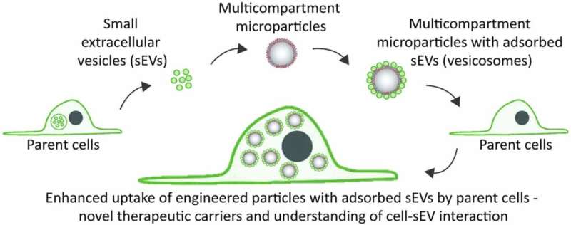
Both Chernyshev and Yashchenok participated in the third study appearing in Colloids and Surfaces B: Biointerfaces, also featuring former Skoltech Ph.D. student Daniil Nozdriukhin and MIPT researchers, which introduced a technique for covering fairly large particles with EVs for the purposes of drug delivery. The team showed that EV-coated particles are readily absorbed by cells, so the approach can be used to feed disguised drug molecules to cells. This would also work with tiny EV-covered sensors, which could be introduced into the cell to explore its inner workings.
According to the teams, further research in collaboration with leading medical centers will involve isolating extracellular vesicles from tumor biopsy samples or blood plasma of actual patients. Another promising line of work is developing bioanalytic platforms for EV extraction and study using microfluidics.
More information: Vasiliy S. Chernyshev et al, Asymmetric depth‐filtration: A versatile and scalable method for high‐yield isolation of extracellular vesicles with low contamination, Journal of Extracellular Vesicles (2022). DOI: 10.1002/jev2.12256
Alexey M. Yashchenok et al, Anti‐CD63‐Oligonucleotide Functionalized Magnetic Beads for the Rapid Isolation of Small Extracellular Vesicles and Detection of EpCAM and HER2 Membrane Receptors using DARPin Probes, Analysis & Sensing (2022). DOI: 10.1002/anse.202200059
Vasiliy S. Chernyshev et al, Engineered multicompartment vesicosomes for selective uptake by living cells, Colloids and Surfaces B: Biointerfaces (2022). DOI: 10.1016/j.colsurfb.2022.112953
Provided by Skolkovo Institute of Science and Technology


