X-ray ptychography performed for first time at small-scale laboratory

In recent years, X-ray ptychography has revolutionized nanoscale phase contrast imaging at large-scale synchrotron sources. The technique produces quantitative phase images with the highest possible spatial resolutions (10's nm) – going well beyond the conventional limitations of the available X-ray optics—and has wide reaching applications across the physical and life sciences. A paper published in Physical Review Letters on 12 May 2021, reveals that an international collaboration of scientists has demonstrated for the first time how the technique of high-resolution phase contrast diffraction imaging can be performed with small-scale laboratory sources.
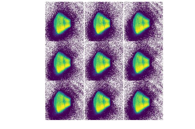
The team from Diamond Light Source, Ghent University, University of Sheffield, and University College London conducted an experiment with a compact liquid metal-jet (LMJ) X-ray source. Laboratory X-ray sources have significantly lower levels of brilliance but currently provide the X-ray synchrotron user community with access to micro-CT, where they can gain a great deal of experience and produce preliminary data, at their home institutions. Until now, no such equivalent has existed for nano-scale imaging through coherent diffraction imaging and ptychography. The team's paper outlines such an experiment and the first proof of concept for far field X-ray ptychography performed using an X-ray laboratory source.
Diamond science group leader, Paul Quinn comments "We have been leading developments in ptychography to open this technique to new science areas and communities. It builds on the work we've done over many years and, longer term this approach in particular has real potential to provide higher resolution imaging to lab source facilities."

The lead author, Darren Batey, Beamline Scientist on the I13-1 Coherence Beamline at Diamond explains Diamond's involvement in this work: "The success of the project strongly depended on the experience and knowledge we collected over the years at the synchrotron. The result of our most recent work allows preliminary studies to be made at Universities, increasing the influx of interesting science to our facility. A lab source facility like the one we have demonstrated with our collaborators, will complement capabilities at Diamond and other sources."
Adding: "Given the worldwide effort toward developing compact light sources, this experimental breakthrough is timely and has the potential of being applied to a whole range of compact light source configurations. The work unlocks the analytical power of ptychography to the wider scientific community and will drive forward the development of advanced coherent diffraction imaging methods. The work with our collaborators ensures that we keep on top of new technology and developments which can enhance the efficiency of experiments at Synchrotron facilities."
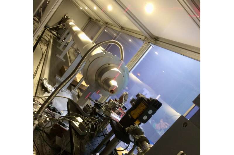
The data were collected at the University of Sheffield Soft Matter AnalyticaL Laboratory (SMALL) with the portable ptychography end-station from I13-1 of Diamond Light Source and a hyperspectral detector from Ghent University. The X-ray source is an Excillum liquid gallium metal jet (LMJ), which has a brilliance of one order of magnitude higher than conventional microfocus sources.
"The resolution achieved in this first experiment is comparable to other lab-based phase contrast techniques, such as in-line phase contrast and edge illumination. The experimental breakthrough achieved with a LMJ is a first step toward expanding X-ray ptychography to other bright compact light sources: from inverse Compton scattering, to laser-plasma based and compact storage rings," says Darren Batey.
More information: X-ray ptychography with a laboratory source. Physical Review Letters, journals.aps.org/prl/accepted/ … c71bef44ba6d228aa184
Journal information: Physical Review Letters
Provided by Diamond Light Source


