Fluorescence microscopy at highest spatial and temporal resolution

LMU researchers simplify the MINFLUX microscope and have succeeded in differentiating molecules that are extremely close together and tracking their dynamics.
Only a few years ago, an ostensibly fundamental resolution limit in optical microscopy was superseded—a breakthrough which in 2014 led to the Nobel Prize in Chemistry for super-resolution microscopy. Since then, there has been another quantum leap in this area, which has further reduced the resolution limit to the molecular level (1 nm).
Scientists at LMU Munich and the University of Buenos Aires have now succeeded in discriminating between molecules that are extremely close together and even tracking their dynamics independently of one another.
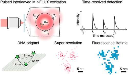
This was achieved by the new p-MINFLUX method by refining and simplifying the recently developed MINFLUX microscope required for 1 nm resolution. Additional functions also permit to distinguish the types of molecules observed. The p-MINFLUX method queries the location of each fluorescently labeled molecule by placing a laser focus close to the molecule. The fluorescence intensity serves as a measure for the distance between the molecule and the center of the laser focus. The exact position of the molecule can then be obtained via triangulation by systematically altering the center of the laser focus relative to the molecule.

The groups led by Professor Philip Tinnefeld (LMU) and Professor Fernando Stefani (Buenos Aires) intercalated the laser pulses in time so that they could switch between the focal positions at the maximum possible speed. In addition, by using fast electronics, a temporal resolution in the range of picoseconds was achieved, which corresponds to electronic transitions within the molecules. In other words, the limits of the microscope are determined exclusively by the fluorescence properties of the dyes used.
In the present publication, the scientists succeeded in showing that the new p-MINFLUX method enables the local distribution of the fluorescence lifetime—the most important measured variable to characterize the environment of dyes—with a resolution of 1 nm. Philip Tinnefeld explains: "With p-MINFLUX it will be possible to uncover structures and dynamics at the molecular level that are fundamental for our understanding of energy transfer processes up to biomolecular reactions."
This project was funded by the German Research Foundation (Cluster of Excellence e-conversion, SFB1032), the Council for Scientific and Technological Research (CONICET) and the National Agency for the Promotion of Research, Technological Development and Innovation (ANPCYT) in Argentina. Prof. Stefani is the Georg Forster Prize winner of the Alexander von Humboldt Foundation and, in this role, a regular guest scientist in physical chemistry at LMU Munich.
More information: Luciano A. Masullo et al. Pulsed Interleaved MINFLUX, Nano Letters (2020). DOI: 10.1021/acs.nanolett.0c04600
Journal information: Nano Letters
Provided by Ludwig Maximilian University of Munich


