Breaking the blur barrier: Working around super-resolution imaging's glitches

Medical researchers face a hurdle when studying cells under an optical microscope—the laws of physics. Obtaining an image of anything below a certain size is complicated; optical apertures and the wavelength of visible light play havoc with clarity. Known as the diffraction limit, it was first encountered by German physicist Ernst Abbe in 1873, and limits the resolution to 200 nanometers (nm) at best (or 200 billionths of a meter).
In the past 20 years, new 'super-resolution' techniques have pushed past this obstacle, imaging items down to few nanometers. One of them, STED (or stimulated emission depletion) microscopy, even won the 2014 Nobel Prize in Physics. But super-resolution has limitations: it either needs complex tools or extensive computer processing, which can add blurry glitches. And it often employs molecular dyes as fluorescent tags, which easily degrade under laser light, making them impossible to use for long exposures.
At the Centre for Nanoscale BioPhotonics (CNBP), scientists are exploring a new strategy that extends the time researchers have to analyze cells under a microscope. It relies on a clever use of a different type of fluorescent marker known as up-conversion nanoparticles, or UCNPs.
"The optical properties of UCNPs give many opportunities for bio-sensing applications and, specifically, for super-resolution imaging," said Dr. Simone De Camillis, a postdoctoral research fellow at CNBP's Macquarie University node, who is part of the team led by Prof Jim Piper, chief investigator for the Advanced Detection and Imaging group.
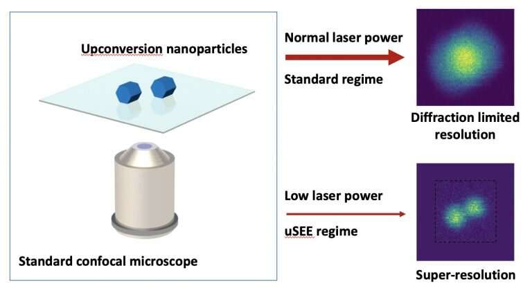
The team developed a new class of UCNPs whose brightness changes abruptly when excited by near-infrared light. This behavior can be exploited to image objects at a resolution half the diffraction limit, so that these extremely small particles can be seen much more clearly. And what's more, the method can be applied to standard confocal microscopes widely used in today's labs.
Because it relies on relatively low-power light, the technique—known as up-conversion super-linear excitation-emission (uSEE) microscopy—is relatively harmless to living cells and could allow imaging deeper into tissue.
The UCNPs can also operate alongside the STED approach, allowing imaging down to 60nm, comparable with the performance of conventional STED using molecular dyes.
The team is now perfecting the design of the new UCNPs and their ability to manufacture them with higher reliability. These improvements, together with improved imaging capability approaching the size of a single nanoparticle, pave the way for 'quantitative imaging': the ability to count the actual number of UCNPs in cells, as well as identify the position of each individual nanoparticle probe and know where they are.
"Currently, when they are very close together, it can be difficult to distinguish them," De Camillis said. "So we are now experimenting with composition and structure of the UCNPs to be able to really resolve single UCNPs, even when they cluster."
Provided by CNBP


