Findings affirm imaging technique's ability to discern healthy tissue after neoadjuvant chemotherapy

In an article published in the peer-reviewed SPIE publication Journal of Biomedical Optics (JBO), titled "Influence of neoadjuvant chemotherapy on diffuse reflectance spectra of tissue in breast surgery specimens," research observed across 92 ex vivo breast specimens suggests that there is little to no impact on the optical signatures of breast tissue after neoadjuvant chemotherapy.
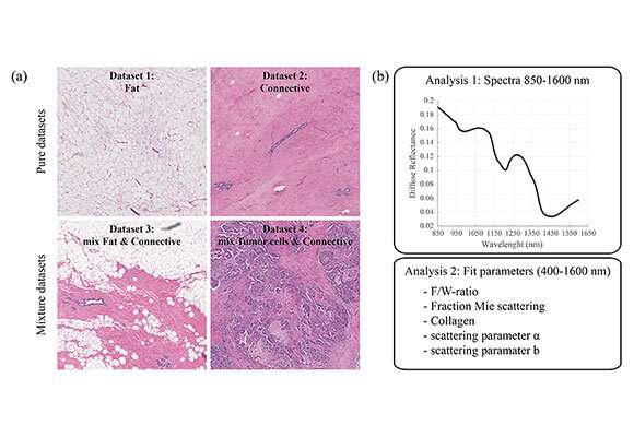
The results of the study, in which diffuse reflectance spectroscopy (DRS) measurements were performed on 92 ex vivo breast specimens from 92 patients treated with and without neoadjuvant chemotherapy show that contrast between healthy tissue and tumor tissue is not altered due to neoadjuvant chemotherapy, suggesting that the same reflectance spectral signatures can be used for tumor margin guidance independent of the chemotherapy status of the patient.
Because healthy and tumor tissue can be readily discriminated, tumor-margin assessment by DRS—which can discriminate different tissue types based on optical characteristics—becomes a feasible consideration during breast-conserving surgery, even if the patient has received neoadjuvant chemotherapy prior to surgery, a procedure that has become commonplace. The ultimate goal of the intra-surgery application of DRS would allow the surgeon to assess the tissue while performing the resection of tumors to ensure that the resection margin is clear of tumor tissue.
According to JBO Editor-in-Chief Brian W. Pogue, the paper and its findings are notable due to the large number of clinical samples analyzed, and the consequent relevance to assessing neoadjuvant chemotherapy changes. "While a significant amount of work has been done defining the spectral signatures of breast cancer tumors and showing that this can be used for guidance, this is one of the first attempts to examine tumors following neoadjuvant chemotherapy as well. The results show that the signatures do not appear to change and so the status of the patient would not confound spectral imaging to help define the lumpectomy margin."
More information: Lisanne L. de Boer et al. Influence of neoadjuvant chemotherapy on diffuse reflectance spectra of tissue in breast surgery specimens, Journal of Biomedical Optics (2019). DOI: 10.1117/1.JBO.24.11.115004
Journal information: Journal of Biomedical Optics
Provided by SPIE


