Noninvasive skull optical clearing window for cortical imaging

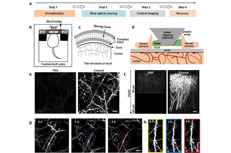
Researchers have demonstrated a noninvasive approach for creating an optical window in the skulls of mice to image their brains. Prof. Dan Zhu and coworkers from Huazhong University of Science and Technology, China, tested the use of optical clearing agents (OCAs) that they applied to the bare skulls (hair and skin removed) of living mice. After treatment with OCAs, the skull becomes transparent within minutes, thus forming a visible window into the cortex. Combined with two-photon microscopy, this technique allows imaging of the fine structures of neurons, glia and the microvasculature in the mouse brain. Given its easy handling, safety, repeatability and excellent performance, this method has promise in neuroscience research. The research has been published in journal Light: Science and Applications.
Observation and manipulation of cells in the cortex is critical to studies of brain structure and function. However, the strong scattering caused by the skull over the cortex limits the penetration depth of light in tissues, and thus hinders the observation of fluorescently labeled neuronal structures and microvasculature. To overcome this obstacle, researcher developed various cranial window methods, including the open-skull glass window, the thinned-skull cranial window, and variants. But these methods present limitations. The tissue optical clearing technique can reduce the scattering of tissues, which has become an important tool for the applications of optical imaging in biomedical research. However, the current optical clearing method is widely used in the ex vivo studies of tissues and organs, and there are few studies on making living tissues transparent.
Prof. Dan Zhu firstly proposed the study of the in vivo optical clearing technique. In the early stage, she was focused on researching different types of skin tissue. Prof. Tonghui Xu, Prof. Dan Zhu's colleague, has been engaged in the research of cortical neuroimaging in mice, and for in vivo cortical imaging, the turbid skull becomes a great bottleneck. After communicating with Prof. Tonghui Xu, Prof. Dan Zhu began to research optical clearing of skull tissue. After six years of hard work, they developed an effective, safe and switchable skull optical clearing window. Through this window, the image contrast and imaging depth are significantly improved, and cortical structures can be imaged at synaptic resolution. This technique holds great promise for studies of brain structure and function in physiological or disease states.
More information: Yan-Jie Zhao et al, Skull optical clearing window for in vivo imaging of the mouse cortex at synaptic resolution, Light: Science & Applications (2018). DOI: 10.1038/lsa.2017.153
Journal information: Light: Science & Applications
Provided by Changchun Institute of Optics, Fine Mechanics and Physics


