Aberrant hyphae triggered by host immune responses to plant pathogenic fungus

Ascochyta (Mycosphaerella) pea blight, caused by Mycosphaerella pinodes, is one of the most important diseases of grain legumes worldwide. Despite the economic impact and numerous studies on this disease, little is known about the cytological features during infection, especially in resistant interactions. One reason is the lack of resistant cultivars of pea, as well as the available resources in the Pisum germplasm collection with strong resistance to this disease.
Kazuhiro Toyoda and colleagues at Okayama University examined the histology and ultrastructure of early infection events and fungal development, including penetration by appressoria, vegetative growth of infection hyphae and host responses, using a recently developed model pathosystem involving Medicago truncatula and M. pinodes strain OMP-1 (Toyoda et al., 2013).
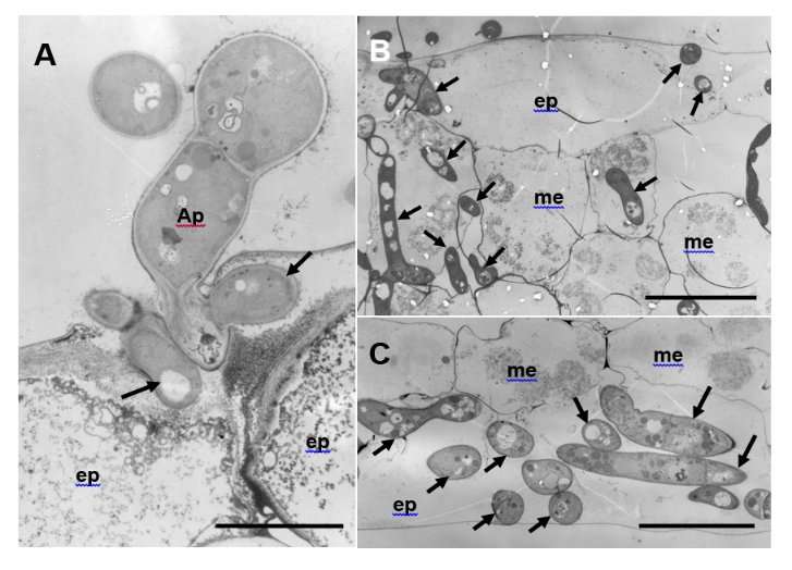
On the susceptible ecotype R108-1, pycnospores germinated and grew over the surface of the epidermis, then formed an appressoria and penetrated the cuticle. Beneath the cuticle, the infection peg expanded into a hyphae that grew within the outer wall of the epidermis. Subsequently, the hyphae penetrated down within mesophyll cells and proliferated vigorously, eventually forming asexual fruiting bodies (pycnidia) (Fig. 1).
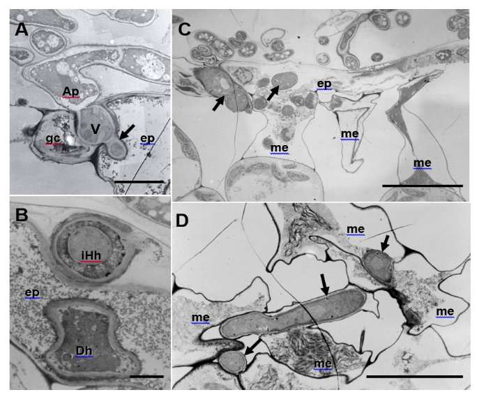
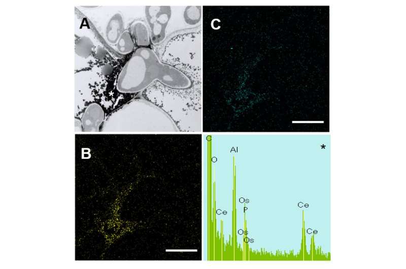
In contrast, successful penetration and subsequent growth of infection hyphae were considerably restricted in the ecotype Caliph (Fig. 2). Interestingly, aberrant hyphae such as intrahyphal hyphae and dead hyphae, due to a local defense elicited by the fungus, were abundant in Caliph but not in R108-1. Detected by its reaction with cerium chloride (CeCl3) to generate electron-dense cerium perhydroxides in transmission electron micrographs, hydrogen peroxide (H2O2) accumulated in epidermal and mesophyll cells of Caliph challenged with pycnospores of M. pinodes. This intracellular localization was confirmed by energy-dispersive X-ray (EDX) spectroscopy (Fig. 3). These observations thus indicate that the oxidative burst reaction leading to the generation of reactive oxygen species is associated with a local host defense response in Caliph, since no clear H2O2 accumulation was detectable in susceptible R108-1.

The researchers conclude that the structural aberrations are likely common mechanisms of fungi for protection from a hostile environment in a resistant host via enclosure by another hyphae. The structural differences between susceptible and resistant interactions as well as the host responses will assist in better understanding pathogenesis of the fungus on pea, thus providing information on the breeding of the resistant cultivars of pea.

More information: Tomoko Suzuki et al. Ultrastructural and Cytological Studies on Mycosphaerella pinodes Infection of the Model Legume Medicago truncatula, Frontiers in Plant Science (2017). DOI: 10.3389/fpls.2017.01132
Provided by Okayama University


