June 19, 2017 feature
Of wrinkles and wires: Capillarity-induced skin folding spontaneously forms aligned DNA nanowire

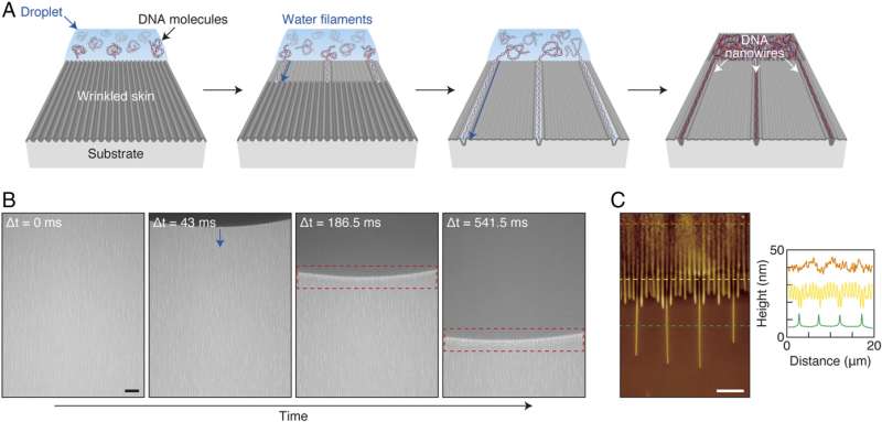
(Phys.org)—Nanowires fashioned from DNA (deoxyribonucleic acid)—one of several type of molecular nanowires incorporating repeating molecular units—are exactly that: Geometrically wire-like DNA-based nanostructures defined variously as having a 1~10 nm (10−9 m) diameter or a length-to-diameter ratio >1000. While nanowires can be made from several organic and inorganic materials, DNA nanowires have been shown to provide a range of valuable applications in programmed self-assembly1,2 of functional materials—including metallic and semiconductor nanowires for use in electronic devices—as well as biological, medical, and genetic analysis applications3,4,5. That being said, DNA nanowire adoption has been limited due to historical limitations in the ability to control their structural parameters—specifically, size, geometry and alignment. Recently, however, scientists at Korea Institute of Science and Technology and Princeton University leveraged the capillary forces of water containing DNA molecules to demonstrate size-controllable straight or undulated aligned DNA nanowires that were spontaneously formed by water entering wrinkled channels of a compressed thin skin on a soft substrate, which subsequently induced a wrinkle-to-fold transition.
Assistant Professor and lead author So Nagashima, Assistant Professor Andrej Košmrlj, Donald R. Dixon '69 and Elizabeth W. Dixon Professor Howard A. Stone, and Principal Research Scientist Myoung-Woon Moon discussed the paper they and their co-authors published in Proceedings of the National Academy of Sciences. "I think that the most challenging aspect of devising our method for utilizing a thin skin template that responds to water by dynamically changing its surface morphology was finding the conditions where the wrinkle-to-fold transition occurs," Moon tells Phys.org. "The critical conditions as a function of the applied strain, initial wrinkle geometries, and thickness of the skin layer determined by oxygen plasma treatment duration were difficult to find." Moon adds that the observation technique for the dynamic transition was limited to only optical microscopes whose highest optical resolution falls between 100 to 1000 nm in the width of nanowires, this being due to the dynamic transition taking place at the submicron scale.
When inducing a template surface wrinkle-to-fold transition by exploiting the capillary forces of water containing DNA molecules, Stone points out, the observation that water changes the wrinkle-to-fold transition is new. "As far as we know, ours is the first study to show this effect, as is demonstrating one use of such folds for the alignment of DNA. Moreover, control of surface tension or resultant capillary forces and the area for fold formation is relatively hard—and by adding DNA molecules to water, it appears that the surface tension is changed, so the fold transition length was shorter."
Template preparation used an oxygen plasma treatment of prestretched polydimethylsiloxane (or PDMS, a polymeric organosilicon compound) substrates for varying durations. "In fact," Moon explains, "the manipulation of PDMS with prestretching strain is a relatively well-developed method as is the oxygen plasma treatment: both have been discussed in the literature. We can make the samples with various sizes of a few millimeters to a few centimeters, which can be also made on much larger area." Moon notes that the researchers can also vary polydimethylsiloxane's mechanical properties—to make it more stretchable, soft or flexible—by changing the ratio of elastomer and cross-linker for PDMS preparation.
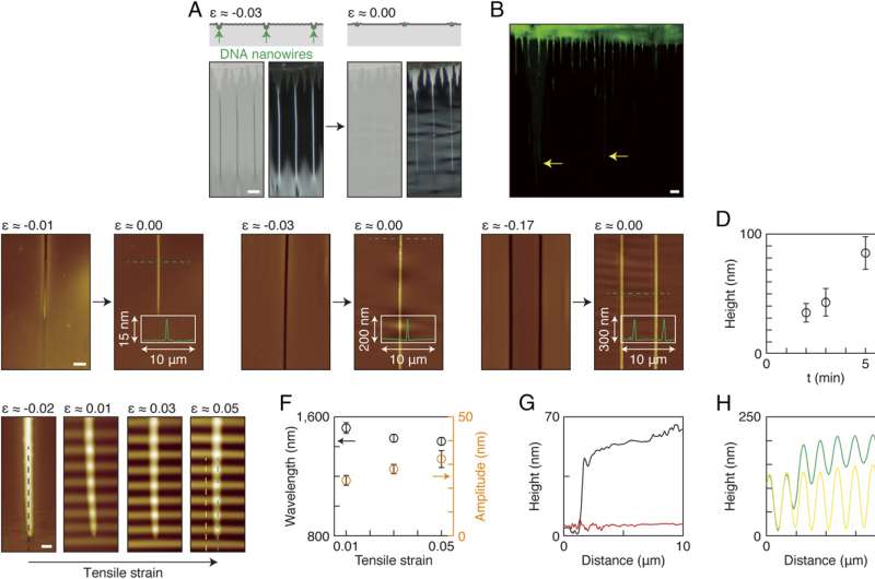
A key aspect of the study was confirming that the new method reliably manipulates DNA nanowire size, geometry, and alignment. "By adjusting the conditions for stretching strain, plasma treatment duration, and post-compression of the stretched PDMS, DNA nanowires can be a half cylinder, a perfect cylinder, or undulated wire shape," Moon tells Phys.org. "By changing the wrinkle geometries such as the amplitude—which is governed by the strain—one can control the distance between wires in the fold channel." Wider distances between wires, he continues, can be accomplished by compressing the PDMS less, while compressing the substrate more yields smaller distances.
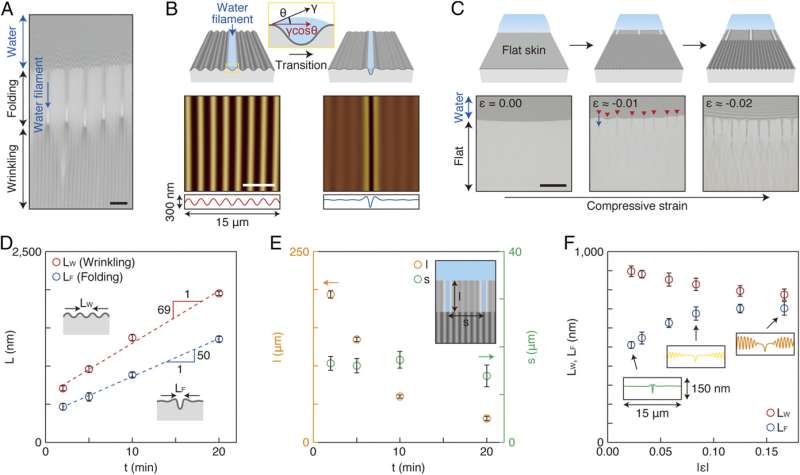
To address these challenges, the scientists discovered a transformation resulting from capillary forces that act at the edge of a water droplet that can, with only 1% compression, transform wrinkles into folds, which in the absence of a liquid drop form only at very high (~30%) compression. In addition, Moon adds, smaller substances such as biomolecules or nanoparticles can follow the water channel to form aligned 1-dimensional nanostructures. "Smaller is better. Less is more. We've found that the wrinkle-to-fold transition takes place more easily when the following factors become smaller: compression level, skin thickness, droplet volume, size of the sample surface, and static contact angles of droplets."
Based on their findings, the authors stated that their approach could lead to new ways of fabricating functional materials. "Our key finding is that one can change wrinkles into localized folds by simply exploiting the capillary forces of water on wrinkled surfaces under very small strain of about 1% in compression," Nagashima tells Phys.org. "Recent studies reported in the literature have demonstrated that such wrinkle-to-fold transitions can help develop systems that dynamically change their properties according to the surface morphology. However, inducing the transition in the absence of water is difficult to achieve in practice because, in general, large compression needs to be applied to the skin-substrate system, which hinders wider applications. Our study reveals that even 1% of compression, which is the critical level for creating wrinkles in our case, is large enough to trigger the transition to folds locally when water is present." Nagashima notes that while the compression level required to induce the transition might differ according to the skin-film system used, only a small compression level would be necessary in combination with water.

"This phenomenon can be considered a lithography-free method that allows for ready fabrication of arrays of nanomaterials, where their size, length, and periodicity could robustly be tuned," he continues. "Moreover, not only water but other liquids could be used to carry nanomaterials and to induce the wrinkle-to-fold transition."
Moon describes several examples of potential de novo fabrication and analysis techniques, including nanoscale lithography, nanoimprint, growth by chemical vapor deposition, and chemical reaction. "Our method can potentially be used for the fabrication of 1-dimensional nanowires or nanoarrays for application to DNA analysis with very dilute or small amounts of DNA; DNA templates as new metal or ceramic nanostructures; and DNA treatment devices for healing modified DNA. In addition, one can adopt this technique to handle protein, blood, or nanoparticles at nanoscale."
Košmrlj and Stone tell Phys.org that one area of planned research is focused on nonlinear analysis and modeling for improved quantitative understanding of the capillarity-induced wrinkle-to-fold transition. "Since our system is composed of the mechanical behavior of the fold transition triggered by liquid surface tension, the wrinkle-to-fold transition that we've found is associated with large deformations where conventional linear elasticity theory does not apply. While the basic mechanisms can be explained within the linear theory, quantitative comparison with experiments can only be achieved by taking into account geometrical and material nonlinearities. We are therefore performing numerical simulations by coupling liquid surface tension and solid deformation, as well as performing analysis with perturbation series, where nonlinearities of elastic structures can be studied systematically."
"I also think that the challenges ahead are to find how to achieve larger areas for DNA pattern formation," Moon says. "In fact, our latest results—obtained after this PNAS article was accepted—shows some impressive progress for the region with wrinkle-to-fold transition in larger areas, such as the entire area underneath a water droplet. Another area to be studied, Moon continues, concerns the fact that biological morphogenesis of skin–substrate systems are ubiquitous in organisms where water is a major constituent. "We're trying to find situations where our findings are applicable. Active collaborations with experts in the field would be helpful."
The researchers might also investigate materials other than PDMS. "Yes. other polymers can work if they possess the basic factors to govern the fold transition, these being the thinness of the nano-skin and soft body materials, and surface hydrophilicity to ensure sufficient surface reaction with liquid," Moon notes.

Other possible future research interests and additional innovations mentioned by the authors include:
- theoretical analysis to elucidate the underlying physics related to the water-induced surface folding
- exploit the underlying physics to develop a robust and mass fabrication method for inducing the wrinkle-to-fold transition
- find and discuss morphological changes in nature where water is likely a key factor
- apply the current study's results to DNA analysis or DNA drug devices
- 2-D/3-D sensors, diagnostic tools, and drug-release systems
- templates for fabricating 1-dimensional nanomaterials
- methods for local patterning
"I believe that this work is beneficial to materials science for nanowire templates, mechanics for fluidic channels, and biology for quantitative analysis of DNA or other biomolecules," Moon concludes.
More information: Spontaneous formation of aligned DNA nanowires by capillarity-induced skin folding, PNAS (2017) 114:24 6233-6237, doi:10.1073/pnas.1700003114
Related:
1DNA nanowire fabrication, Nanotechnology (2006) 17:R14m https://core.ac.uk/download/pdf/1559975.pdf
2DNA-Templated Self-Assembly of Protein Arrays and Highly Conductive Nanowires, Science (2003) 301:5641 1882-1884, doi:10.1126/science.1089389
3Nanowire-Based Sensors for Biological and Medical Applications, IEEE Transactions on NanoBioscience (2016) 15:3 186-199, doi:10.1109/TNB.2016.2528258
4DNA-Based Applications in Nanobiotechnology, Journal of Biomedicine and Biotechnology (2010) Article ID 715295, doi:10.1155/2010/715295
5Nanowire nanosensors, Materials Today (2005) 8:5, doi:10.1016/S1369-7021(05)00791-1
Journal information: Proceedings of the National Academy of Sciences , Nanotechnology , Science , Journal of Biomedicine and Biotechnology
© 2017 Phys.org


