Fungus a possible precursor of severe respiratory diseases in pigs

Respiratory diseases in pigs typically involve multiple infections from different pathogens. Some pathogens play a greater role than others in the progression of the disease. The fungus Pneumocystis carinii is a relatively common cause of pneumonia in Austrian pigs, but its role has so far remained largely unexplored. Pneumocystis is considered to be less dangerous than other pathogens, as it probably requires other underlying conditions to sufficiently weaken the immune defence of the animals first.
A research team from Vetmeduni Vienna has now demonstrated the susceptibility of piglets to the fungus as well as its relation with other pathogens in the progression of the disease. Their research has shown that the fungus plays a more important role in pneumonia than had previously been assumed. Detecting pneumocystis in a medical examination requires a lung lavage, which, however, is less stressful to the animals than other sampling methods.
Pneumocystis in sick piglets of all age groups
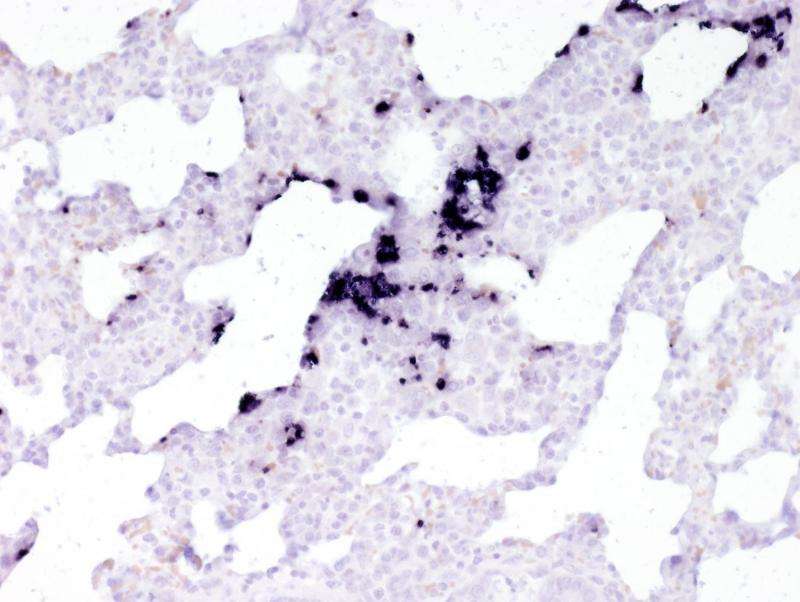
The researchers began by testing stored tissue samples of sick piglets for the presence of the fungus and other pathogens. Pneumocystis was detected in piglets of all ages. In suckling piglets, only the fungus was detected in lung tissue. The older the piglets, the more bacteria could be detected. Pneumocystis itself was no longer as prevalent as in the suckling piglets. The fungus, however, appeared to have proliferated before the bacterial pathogens.
This suggests that the fungus plays a role as a "door opener" for secondary infections. "First the fungus spreads along the alveolar walls. From there it proliferates and fills the alveolar spaces. As a result, the lung tissue receives insufficient oxygen and bacteria can reproduce better in the damaged tissue," explains Christiane Weissenbacher-Lang of the Institute for Pathology and Forensic Veterinary Medicine, describing the possible progression of the infection.
Fungus detectable in living pigs only with lung lavage
The progress of the infection can be easily demonstrated in the laboratory using lung biopsies. In living pigs, however, this type of sampling is difficult and not suitable for routine testing. The team around Weissenbacher-Lang therefore tested whether oral fluid samples and lung lavages from sick pigs could be suitable for a diagnosis. Lung lavages are less stressful and easier to perform than biopsies. They also offer the advantage that material is collected from the entire lung.

Role of Pneumocystis carinii likely underestimated
The researchers demonstrated that it was possible to clearly diagnose even a weak infection with the material from a lavage. The presence of fungal DNA can be specifically demonstrated using molecular methods such as real-time PCR. The test was designed to detect the strains typically found in pigs. Oral fluid samples were determined to be not suitable for diagnosing mild or moderate infections. Sampling through lung lavages therefore appears to be the best method for diagnosing the fungus in routine testing among living pigs.
Weissenbacher-Lang intends to conduct further tests to show that pneumocystis pneumonia can play a role as an additional factor in co-infections with other respiratory pathogens. If this were the case, it would be necessary to respond in time. The high susceptibility demonstrated among the tested pigs shows that pneumocystis should not be underestimated, even if the fungus itself has so far been categorized as a low risk for pigs.
Lung lavage an option for routine testing
Respiratory diseases in pigs are an economic risk for farms. The potential loss is great. Keeping the animals in pens therefore requires high quality standards for the ventilation alone. If a pathogen still manages to manifest itself in the pen, veterinary measures must be taken in time to keep the majority of the herd healthy. Lung lavages are a suitable method for regular testing of the herd.
Biopsies time-consuming and potentially harmful to the animals
Biopsies require the animals to be anaesthetized and samples to be collected under ultrasound guidance. The procedure is time-consuming, stressful for the animal and involves potential health risks. Lung lavages are much less stressful and take less time to perform. The pigs are anaesthetized only briefly. The lavage itself takes only a few minutes. Lavages are a routine method in human medicine. Veterinarians are also familiar with this procedure and the method could therefore be applied in the pen without any major adaptations necessary for testing.
More information: C. Weissenbacher-Lang et al. Establishment of a quantitative real-time PCR for the detection of Pneumocystis carinii f. sp. suis in bronchoalveolar lavage samples from pigs, Journal of Veterinary Diagnostic Investigation (2016). DOI: 10.1177/1040638716641158
B. Kureljušić et al. Association between Pneumocystis spp. and co-infections with Bordetella bronchiseptica, Mycoplasma hyopneumoniae and Pasteurella multocida in Austrian pigs with pneumonia, The Veterinary Journal (2016). DOI: 10.1016/j.tvjl.2015.11.003
Provided by University of Veterinary Medicine—Vienna


