Novel microscopy method illuminates cell changes caused by aging, injury and disease
A University of Maryland-led team of researchers has developed an optical microscopy technique capable of shedding new light on how the mechanical properties of cells change in the course of aging, injury healing and disease pathogenesis.
The technique offers promise that one day researchers will be able to identify a more exact starting point for the development of cancers, coronary disease or even osteoporosis. "Using light, we can measure cells inside tissue and we can look inside the cells to distinguish, for example, the properties of the nucleus and cytoplasm," said team lead Giuliano Scarcelli, assistant professor with UMD's Fischell Department of Bioengineering.
The team of researchers from UMD, the Massachusetts Institute of Technology (MIT) and Massachusetts General Hospital published their work this week in Nature Methods.
Scientists need a clearer look at how properties of cells change over time, both to learn more about factors that influence everyday biological functions of cells and to better understand how diseases develop.
Every cell contains a cytoplasm – the thick solution enclosed within the cell by the cell membrane. Primarily composed of water, salts, and proteins, the cytoplasm is where most cellular activities occur. Even more, the cytoplasm serves as a means of transport for genetic material and acts as a buffer, protecting the cell's genetic material from damage due to movement or collision with other cells.
For years, researchers have known that the interaction between the liquid and solid phases within the cytoplasm plays a prominent role in how cells deform and move. As such, the ability to map the hydro-mechanical properties of cells – such as viscoelasticity and compressibility – is critical to advancing understanding of how cell properties change as a symptom of disease in the body or as part of normal biological functions, such as when wounds heal.
Traditionally, techniques used to study the mechanical properties of cells have either required contact with cells or have produced images with limited resolution. As a result, information on the biomechanical properties of cells in 3-D environments is lacking.
"Gold-standard techniques require contact, therefore they are inherently limited to providing global averages of cell properties and to experimental situations where you have physical access to the cell," Scarcelli said.
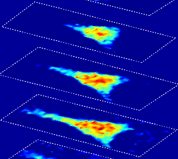
To address this, Scarcelli and colleagues from the Massachusetts Institute of Technology (MIT), and Massachusetts General Hospital have introduced a technique known as Brillouin optical cell microscopy for noninvasive, 3-D mapping of intracellular and extracellular hydro-mechanical properties. Their technique employs Brillouin light scattering, a process that occurs when light interacts with density fluctuations in a medium. These spontaneous fluctuations are driven by collective acoustic vibrational modes, known as phonons, in the gigahertz frequency range. In this way, Brillouin microscopy yields invaluable information on the viscoelastic characteristics of cells – and does so at a microscopic resolution no other technique can match.
As such, Brillouin microscopy opens up new research avenues for the biomechanical investigation of cells and their microenvironment in 3-D at subcellular resolution.
Continuing in collaboration with Roger Kamm at MIT, the research team is now taking advantage of the unique capabilities of Brillouin microscopy to look at a crucial property of metastatic cells – their ability to modulate their internal mechanical properties to enter and exit blood vessels to colonize distant sites. The team's efforts recently earned them a five-year, $3 million National Institutes of Health grant to study tumor cell extravasation.
More information: "Noncontact three-dimensional mapping of intracellular hydro-mechanical properties by Brillouin microscopy." Nature Methods DOI: 10.1038/nmeth.3616
Journal information: Nature Methods
Provided by University of Maryland


