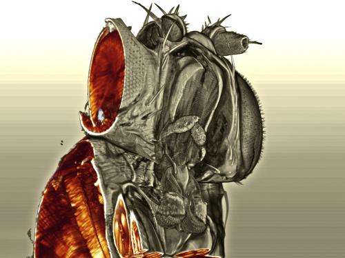
Video of the inside of a fly's eye: Scientists develop laser optics which support high-resolution 3D microscopy

Ultra-microscopes developed at the Vienna University of Technology can look into biological tissue, creating high-resolution 3-D images. A video has been created, showing a 3D-scan through a fly's head.
Fine venules, thin branches of nerve tracts - thanks to the ultramicroscope developed at the Bioelectronics Department of the Institute for Solid-State Electronics at the Vienna University of Technology, the tiniest details of biological tissues can be represented in 3D. Laser beams are used to look inside flies, mice, or medical tissue samples. The laser technology and the optics in the device were developed by Saideh Saghafi. Using various optical tricks, she has managed to turn a laser beam into an extremely thin two-dimensional laser surface, which can be shone through samples layer by layer. She has now been awarded a major optics prize for this work.
Tissue made transparent
Biological tissue tends to be opaque, with light being scattered at the interfaces between different materials. This is why we cannot see through thick fog: each individual floating droplet of fog scatters the light, so all we can see is a blanket of white.
In order to represent the internal structure of biological tissue, it must first be made transparent to laser beams. 'The sample is treated first of all: any water it contains is replaced with a fluid with different optical properties, and this enables laser beams to penetrate deep into the sample,' explains Saideh Saghafi. Together with her colleagues at the department of Prof. Hans Ulrich Dodt at the Vienna University of Technology, she is creating images of previously unmatched quality, which are providing important information for medical research. The novel ultramicroscope is also ideal for the investigation and 3D representation of human tumours from a pathology perspective.
Ultra-thin light surfaces
Optical tricks are initially used to convert a conventional round laser beam into an elliptical beam, which is transformed in turn into a thin layer of light. 'The surface of the laser light, which we generate with our lenses, is only around 1.5 micrometres thick,' says Saghafi. Stimulated by the laser light, an extremely thin layer of the sample begins to fluoresce - and this light can be picked up with a camera. The basic idea behind ultramicroscopy has been applied at the Vienna University of Technology for some years now, but Saghafi's thin laser layers have made a further decisive improvement in terms of microscope precision.
Laser light is shone through the sample layer by layer, with an image being taken each time. These are used to construct a complete 3D model of the sample on the computer. Detailed images emerge of tiny fruit files and the complex network of neurons in the brains of mice. 'If we didn't shine the laser surface through the sample, it would be a case of having to cut the sample into thin layers and then put these under a microscope one at a time. Of course, this approach could never match the accuracy we achieve with our ultramicroscope,' explains Saideh Saghafi.
Prize for outstanding scientific achievement
Edmund Optics, a major manufacturer of optical equipment, recently presented a series of awards for the best scientific work in the field of optics. From the 750 or so entries, the three most innovative and useful from a technical perspective were selected. Saideh Saghafi featured among the work receiving awards for 2012 with her light surface technology. The prize money is paid in the form of valuable optical equipment, which should help ultramicroscopy improve still further as a discipline at the Vienna University of Technology.
Provided by Vienna University of Technology


