Microscope technique reveals for first time when and where proteins are made

Scientists at Albert Einstein College of Medicine of Yeshiva University and their international collaborators have developed a novel fluorescence microscopy technique that for the first time shows where and when proteins are produced. The technique allows researchers to directly observe individual messenger RNA molecules (mRNAs) as they are translated into proteins in living cells. The technique, carried out in living human cells and fruit flies, should help reveal how irregularities in protein synthesis contribute to developmental abnormalities and human disease processes including those involved in Alzheimer's disease and other memory-related disorders.
The research will be published the March 20 edition of Science.
"We've never been able to pinpoint exactly when and where mRNAs are translated into proteins," said study co-leader Robert H. Singer, Ph.D., professor and co-chair of anatomy and structural biology and co-director of the Gruss Lipper Biophotonics Center at Einstein. "This capability will be critical for studying the molecular basis of disease, for example, how dysregulation of protein synthesis in brain cells can lead to the memory deficits that occur in neurodegeneration." Dr. Singer also holds the Harold and Muriel Block Chair in Anatomy & Structural Biology at Einstein.
The directions for making proteins are encoded in genes in the cell nucleus. Two steps—transcription and translation—must occur so that the gene's protein-making instructions will lead to actual proteins. In the first step, called transcription, the gene's DNA is "read" by molecules of mRNA. These mRNAs then migrate from the nucleus into the cytoplasm and attach to structures called ribosomes. That's where translation, the second step in protein synthesis, occurs: the mRNAs attached to ribosomes function as templates on which proteins are constructed.
To visualize translation, Dr. Singer and his colleagues took advantage of a key occurrence during the first round of translation: the ribosome to which mRNAs attach must displace so-called RNA-binding proteins from the mRNAs. The researchers synthesized identical copies of mRNA molecules containing two fluorescent proteins, one green and one red. This meant that in the nucleus (where mRNAs are made), mRNAs labeled with both red and green proteins appear yellow. After migrating to the cytoplasm, the mRNAs can change color depending on their fate.
For mRNAs landing on ribosomes, the ribosome displaces the mRNAs' green fluorescent protein. As a result, these mRNA molecules—stripped of their green fluorescent proteins, bound to ribosomes, and ready to be translated into a protein—appear red. Meanwhile, all the untranslated mRNA molecules remain yellow. The technique was dubbed TRICK (for Translating RNA Imaging by Coat protein Knock-off).
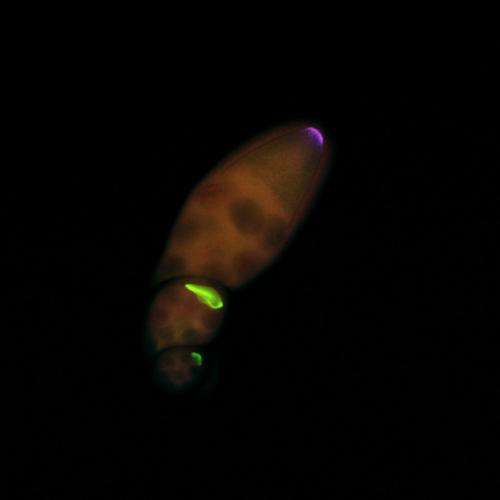
In a test of TRICK, the collaborators in Germany studied when and where mRNAs for a gene called oskar are expressed in Drosophila eggs, or oocytes. (Drosophila, or fruit flies, are a frequently used model for understanding human disease, and oskar is critical for normal development of fruit fly embryos.) The researchers made oskar mRNAs tagged with red and green fluorescent proteins and inserted the tagged mRNAs into the nuclei of Drosophila oocytes.
"Using TRICK, oskar mRNAs were not translated until they reached the posterior pole of the oocyte," said Dr .Singer. "We suspected this, but now we have definitive proof. Going forward, researchers can use this technique to dissect the cascade of regulatory events required for mRNA translation during Drosophila development."
The researchers also found that protein translation doesn't start immediately after mRNAs exit the nucleus but instead gets underway several minutes after mRNAs have entered the cytoplasm. "We never knew that timeframe before," said Dr. Singer. "That's another example of what we can learn using this technique."
More information: An RNA biosensor for imaging the first round of translation from single cells to living animals, Science, www.sciencemag.org/lookup/doi/ … 1126/science.aaa3380
Journal information: Science
Provided by Albert Einstein College of Medicine


