October 9, 2014 report
Improvements to scanning transmission electron microscope now allows vibrational spectroscopy

(Phys.org) —A team of researchers in the U.S. has added the ability to detect atomic lattice vibrations to a scanning tunneling electron microscope. In their paper describing their efforts, published in the journal Nature, the team describes how they applied new advances in vibrational spectroscopy to the electron microscope to allow for better study of such things as nanostructures involved in dynamic processes. Rik Brydson, of the University of Leeds in the U.K. offers a New & Views piece on the work done by the team in the same journal issue.
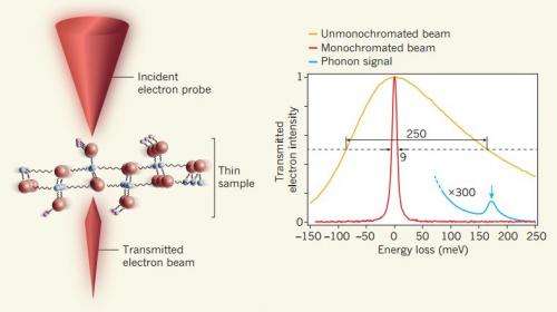
The electron microscope has allowed scientists to see things that until its invention, no one thought possible. Unfortunately, for all its strengths, it's not been able to allow for watching ongoing processes at the atomic level—for that, researchers have had to turn to vibrational spectroscopies such as Ramen scattering, low energy electrons or infrared radiation. Now, because of advances in several technologies, the researchers with this new effort report that they have been able to add the ability to detect atomic lattice vibrations to an electron microscope. To make that happen, they increased the sensitivity when conducting electron energy-loss spectroscopy allowing them to measure how much energy electrons were losing as they moved through a material, which allowed for calculating how much of that energy went into causing vibrations. For the case where electrons move through a material without losing energy, the new device allows for better detection of the peaks in spectra that occur.
The improvement marks a rarity, Brydson notes—where a development in instrumentation delivered an order of magnitude improvement on existing technology, without sacrificing performance in other areas. It also, he adds, represents an excellent example of the research community (a combination of academics and those in the private sector) working together in harmony to produce something truly good and useful.
The new technique opens the door to allowing researchers to watch (or film) processes such as catalysts, transfer of heat, and what happens at a closer level in solar cells as they collect sunlight. And that opens the door to allowing for tweaking such processes in ways that cause them to come about with better outcomes.
More information: Vibrational spectroscopy in the electron microscope, Nature 514, 209–212 (09 October 2014) DOI: 10.1038/nature13870
Abstract
Vibrational spectroscopies using infrared radiation, Raman scattering, neutrons, low-energy electrons and inelastic electron tunnelling6 are powerful techniques that can analyse bonding arrangements, identify chemical compounds and probe many other important properties of materials. The spatial resolution of these spectroscopies is typically one micrometre or more, although it can reach a few tens of nanometres or even a few ångströms when enhanced by the presence of a sharp metallic tip. If vibrational spectroscopy could be combined with the spatial resolution and flexibility of the transmission electron microscope, it would open up the study of vibrational modes in many different types of nanostructures. Unfortunately, the energy resolution of electron energy loss spectroscopy performed in the electron microscope has until now been too poor to allow such a combination. Recent developments that have improved the attainable energy resolution of electron energy loss spectroscopy in a scanning transmission electron microscope to around ten millielectronvolts now allow vibrational spectroscopy to be carried out in the electron microscope. Here we describe the innovations responsible for the progress, and present examples of applications in inorganic and organic materials, including the detection of hydrogen. We also demonstrate that the vibrational signal has both high- and low-spatial-resolution components, that the first component can be used to map vibrational features at nanometre-level resolution, and that the second component can be used for analysis carried out with the beam positioned just outside the sample—that is, for 'aloof' spectroscopy that largely avoids radiation damage.
Journal information: Nature
© 2014 Phys.org


