Ultra-compact implantable image sensor using body channel communication

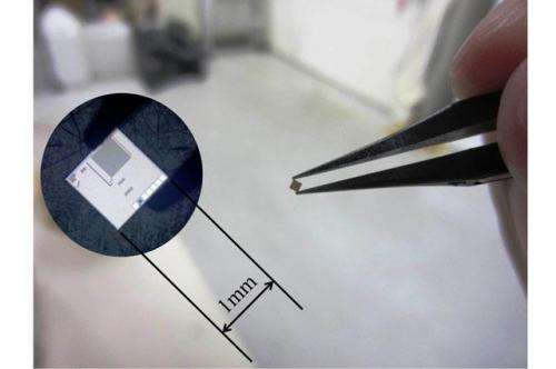
An ultra-compact implantable image sensor using body channel communication has been demonstrated in Japan. The body channel approach allows the sensor-transmitter device to be much smaller and use less power than an RF wireless unit.
Fundamental limits
Interest in implantable medical sensors is on the rise as developments in established technologies and new concepts are making more and more applications feasible. One thing that all such sensors share is the need to be able to get the information they gather within the body, out of the body.
RF communication is widely used for such applications, however, with implantable devices size reduction is generally desirable to reduce invasiveness, and in some applications there are also specific size limitations, owing to where and how the sensors are to be implanted. For example, sensors intended for use inside the brain need to be very compact.
Using smaller antennas generally means using higher frequencies, and that in turn leads to attenuation problems with biological tissues. To compensate for increased attenuation more power is needed, which is also a serious issue for an implantable device.
Conductive communication
An alternative wireless approach to sending the data is body channel communication, in which an electrical signal is transmitted by conduction through the tissues of the body. In this issue of Electronics Letters, researchers from the Nara Institute of Science and Technology's Graduate School of Materials Science report using this approach to transmit and receive image data from a CMOS sensor fully implanted in a simulated body environment.

Their sensor design, using body channel as the transmission method, will allow data to be read from the device by attaching an electrode to the surface of the body near the implantation site, as well as allowing the sensor unit itself to be low-power and small.
"Our implantable CMOS image sensor is intended for biomedical applications such as brain functional imaging," explains team member Hajime Hayami. "It can be planted with minimum invasiveness. Its features enable implantation of a number of sensors in a brain to investigate collaborative neural activities."
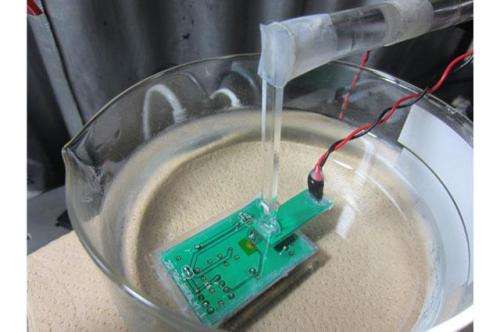
In their experiments, the team at Nara have transmitted images from a CMOS sensor submerged in phosphate buffer saline (PBS), a body simulant material, to a receiver electrode 2 mm away. The experiments prove the principle of operation for this form of communication and even this small distance is enough to allow in vivo use in smaller animals.
"We are planning to apply our device to brain functional imaging of a mouse brain in the near future. Because the size of a mouse brain is only a few mm thick, the transmission distance of 2 mm is sufficient. Even in the case of signal transmission through a longer distance with a larger animal, the experimental results indicate that this method can be applied by adjusting the sensitivity of the receiver circuit," said Hayami.
Neural arrays
Before the team can proceed with implantation in an animal, they need to integrate all of their PCB based devices into a single chip. This chip has already been designed and they are now working on the fabrication, with the goal of beginning implantation experiments by the end of 2014.
Meanwhile, the Nara researchers have also been working to develop the design and say that relatively long distance transmission is now possible through improvements to the receiver circuit. On the sensor/transmitter side, they have also shown that they can use pulse-width modulation rather than an ADC output from the image sensor.
The central goal of their work is creating tools for research into the human brain. "Our research group aims to elucidate the cooperative neural activity with distributed ultra-small sensors in the brain. We think we can achieve the goal by designing more intelligent chips based on the proposed communication method," said Hayami.
The team believe that current sociological trends, including aging societies, will continue to drive demand for implantable devices, and that the applications of this kind of technology may develop to include more natural interaction between users and technology. Hayami commented that "I hope we will create an epoch where one can unconsciously use implantable devices by developing a brain-machine interface."
More information: "Digital signal transmission from fully implantable CMOS image sensor in simulated body environment" H. Hayami, et al. Electronics Letters, Volume 50, Issue 12, 05 June 2014, p. 851 – 853. DOI: 10.1049/el.2014.0765 , Print ISSN 0013-5194, Online ISSN 1350-911X
Journal information: Electronics Letters
Provided by Institution of Engineering and Technology


