Knee-deep sensing

A new, non-invasive technique to track the motion of knee bones in 3D with a very high precision has been presented by researchers in Australia. By employing a single-element ultrasound sensor and a fast image registration technique, the team of collaborating researchers from University of New South Wales, University of Canberra and Canberra Hospital achieved sub-millimetre precision, which is essential for clinical applications such as artificial joint component design and diagnosis for ligament injuries.
A joint effort
The kinematic analysis of the motion of the individual bones in the knee joint gives essential insight into many applications. Orthopaedic surgeons will use this information for knee replacement and reconstruction surgery with the goal of restoring normal motion to the knee joint after a total knee replacement or surgery to repair ruptured ligaments.
Kinematic analysis can also be used in other important applications such as gait analysis, normal and abnormal joint trajectory detection in motion analysis, artificial joint component design and rehabilitation, diagnosis for ligament injuries and therapeutic strategy formulation.
Currently, the clinical standard for performing kinematic analysis with sufficient precision involves implanting tantalum beads into the bones, which then appear as high intensity contrast features in radiographs. This technique, however, has significant disadvantages: it is invasive; the patient has to be exposed to X-rays; and the procedure requires the patient to be lying in a horizontal position so cannot be used to capture the 3D motion analysis of the knee joint during normal activities.

Some alternative solutions do allow the capture of the kinematics during normal activities; the merging of 2D X-ray videos with 3D CT scans is one method, but still has the disadvantages of exposure to radiation and a limited field of view. Other proposed methods have used optoelectronic, ultrasonic or video-based systems to track markers on the skin, but the markers can move independently of the underlying bone, making the technique less precise.
A full range of motion
As part of their work into developing novel techniques to aid in the diagnosis of joint deficiencies and artificial joint component design, the researchers took a different approach to the visual representation of knee kinematics by making use of a commercially available, small and lightweight ultrasound sensor. Placed on the skin above the tibia and femur, it is non-invasive, but can penetrate into the muscle tissue on the knee joints to provide high resolution images of the motion of the bones.
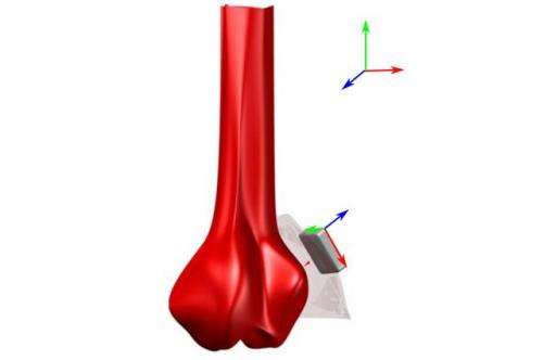
This type of single-element ultrasound sensor can sweep any pre-defined trajectory as it has a single line-of-sight. In their set-up, the researchers selected motion trajectories for the image acquisition so that the acquired 2D image slices could be used to automatically determine the 3D motion parameters in order to model the knee joint kinematics. The researchers developed and coded a 2D/2D image registration technique to achieve this.
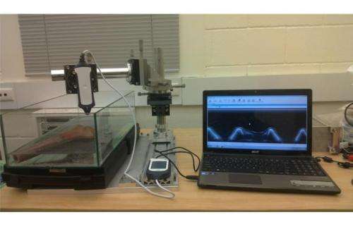
Tests were carried out on a prototype system, primarily consisting of a phantom bone model immersed in a tank of water, and precision micro-mechanical stages which enabled the sensor to be positioned using three translational and three rotational directions. Using their prototype, the researchers proved that ultrasound can provide precision of less than a millimetre and that it was able to capture anatomical details of bones in the knee joints relative to the sensor, making it a suitable technique for the measurement of knee joint kinematics in clinical use.
Future directions
For this system to be realised for clinical applications, the single element sensor with micro-motors has to be miniaturised in an appropriate casing, filled with acoustic impedance matching liquids and interfacing electronics for signal transmission and reception.
The team are currently working on the sensor miniaturisation along with developing software for kinematic data representation in 3D. They are also developing a signal transmission and reception protocol for the sensor using a field programmable gate array.
The technique presented in this Letter, although intended for the motion analysis of knee joints, could be flexible enough to be used for other human joints such as the shoulder or the hip. The researchers hope to see 3D motion parameter estimation from a few 2D projections of the human anatomy becoming more widespread in medical applications.
More information: "Precision analysis of single-element ultrasound sensor for kinematic analysis of knee joints." M.A. Masum, et al. Electronics Letters, Volume 50, Issue 15, 17 July 2014, p. 1047 – 1048. DOI: 10.1049/el.2014.1230 , Print ISSN 0013-5194, Online ISSN 1350-911X
Journal information: Electronics Letters
Provided by Institution of Engineering and Technology


