Microscopy charges ahead

(Phys.org) —Ferroelectric materials – substances in which there is a slight and reversible shift of positive and negative charges – have surfaces that are coated with electrical charges like roads covered in snow. Accumulations can obscure lane markings, making everyone unsure which direction traffic ought to flow; in the case of ferroelectrics, these accumulations are other charges that "screen" the true polarization of different regions of the material.
Ferroelectric materials are of special interest to researchers as a potential new form of computer memory and for sensor technologies.
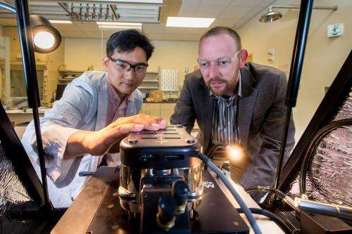
In order to see this true polarization quickly and efficiently, researchers at the U.S. Department of Energy's Argonne National Laboratory have developed a new technique called charge gradient microscopy. Charge gradient microscopy uses the tip of a conventional atomic force microscope to scrape and collect the surface screen charges.
"The whole process works much like a snowplow scraping along the roads," said Argonne materials scientist Seungbum Hong, who led the research. "Before, all we had was a snowshovel."
Ferroelectric materials are not usually polarized in any particular way, but they are rather the combination of different domains that are each polarized in different directions. "The end goal of the research is to be able to map these different regions quickly and accurately," Hong said.
"Until now, the process of trying to map these regions has been incredibly arduous and time-consuming," added Argonne Nanoscience and Technology interim division director Andreas Roelofs, who came up with the idea for the study. "What was taking us 10 to 15 minutes now takes seconds."
Previous efforts in this arena had focused on the application of a different kind of microscope using piezoresponse force microscopy (PFM). In this technique, an applied voltage causes a small displacement of atoms in the material, generating a noticeable mechanical effect, or vibration. In reverse, the same phenomenon is responsible for the workings of the lighters in gas grills.
The problem with PFM is that it is very slow and requires sophisticated equipment to measure a tiny motion of the material. "Before, we had to sit on one spot for a long time to get enough signal to understand how the material moves because we could just barely sense it," Roelofs said. "For the past 15 years or so, we've tried to increase the speed of the measurements and made only modest progress while adding a lot of complexity."
"Now, everyone can use a standard tool to do this work much more cheaply and efficiently," he added.
An article based on the study appears in the April 23 early edition of the Proceedings of the National Academy of Sciences.
More information: Seungbum Hong, Sheng Tong, Woon Ik Park, Yoshiomi Hiranaga, Yasuo Cho, and Andreas Roelofs. "Charge gradient microscopy." PNAS 2014 111 (18) 6566-6569; published ahead of print April 23, 2014, DOI: 10.1073/pnas.1324178111
Journal information: Proceedings of the National Academy of Sciences
Provided by Argonne National Laboratory


