High-resolution microscopy technique resolves individual carbon nanotubes under ambient conditions

Carbon nanotubes are expected to be used in a myriad of applications ranging from military protective clothing to hydrogen storage. Due to their nanometer dimensions, however, the structure and surface chemistry of individual carbon nanotubes cannot be easily studied using conventional techniques. Norihiko Hayazawa and colleagues from the Near Field NanoPhotonics Research Team at the RIKEN Center for Advanced Photonics have now developed a high-resolution microscopy technique that can resolve individual carbon nanotubes under ambient conditions.
Raman spectroscopy is widely used to probe the characteristics of materials with high precision. It involves exciting the material surface with a laser and then measuring the change in laser energy after it is scattered from the surface. Tip-enhanced Raman spectroscopy (TERS) is used to achieve close to molecular resolution by passing a metallic tip across the material surface to enhance the Raman signals of nearby molecules. TERS using an atomic force microscope (AFM) tip is capable of simultaneously assessing the structure and surface chemistry of materials at a resolution of around 10–20 nanometers—far below the diffraction limit of conventional optical microscopes.
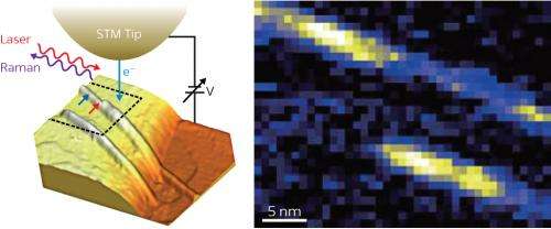
Replacing the AFM tip with a scanning tunneling microscope (STM) tip has recently been shown to improve the resolution of the technique dramatically. The position of the metallic STM tip can be controlled more precisely than that of an AFM, making it possible to scan a material with a gap between the tip and surface of less than 1 nanometer. Strong coupling between electronic resonances called 'plasmons' of the tip and material surface across this narrow gap in STM-TERS further improves the resolution of the technique (Fig. 1).
"With our STM-TERS system, we have achieved resolution of 1.7 nanometers, meaning that carbon nanotubes can be visualized at the dimensions of their diameter," explains Hayazawa. "This makes it possible for the first time to extract the local property of the carbon nanotubes optically without averaging."
Whereas previous nanoscale STM-based techniques and STM-TERS methods have required cryogenic temperatures and ultrahigh vacuums, the STM-TERS technique developed by Hayazawa's team can be used with a compact chamber at ambient pressure and temperature. This significantly broadens the range of materials that can be probed. "DNA sequencing, protein dynamics on biological membranes, and organic solar cells all require ambient conditions," explains Hayazawa.
As well as using the technique to probe other materials at ultrahigh resolution, the researchers hope to be able to reveal previously undiscovered physical properties of carbon nanotubes.
More information: Chen, C., Hayazawa, N. & Kawata, S. A 1.7 nm resolution chemical analysis of carbon nanotubes by tip-enhanced Raman imaging in the ambient. Nature Communications 5, 3312 (2014). DOI: 10.1038/ncomms4312
Journal information: Nature Communications
Provided by RIKEN


