This article has been reviewed according to Science X's editorial process and policies. Editors have highlighted the following attributes while ensuring the content's credibility:
fact-checked
peer-reviewed publication
proofread
Novel time-of-flight-resolved stimulated Raman scattering microscopy enables high-resolution bioimaging

Stimulated Raman scattering (SRS) microscopy is an optical vibrational spectroscopic imaging technique and has emerged as an appealing label-free imaging tool for tissue and cell imaging and characterization with high biochemical specificity.
However, the tightly focused Gaussian excitation beams used in conventional SRS microscopy experience a strong light scattering effect that deteriorates the beam profile during propagation into turbid media due to inhomogeneous refractive indices in tissue, leading to a degraded spatial resolution and limited light penetration incapable of volumetric deep tissue imaging.
The use of non-diffracting Bessel beams has appeared as a promising alternative to improving penetration depth for 3D deep imaging, but raster scanning with Bessel beams can only provide SRS 2D projection images of the sample and thus, the depth information is lost.
Additionally, existing Bessel beam-based SRS tomography relies on optical beating or optical projection techniques to encode the depth information spatially with acquisition of multiple raw images, which might suffer from sample motion artifacts.
In a new paper published in Light: Science & Applications, a team of scientists, led by Professor Zhiwei Huang from Optical Bioimaging Laboratory in the Department of Biomedical Engineering, College of Design and Engineering, National University of Singapore, Singapore, have developed a novel time-of-flight resolved Bessel light bullet-enabled stimulated Raman scattering (B2-SRS) microscopy for deeper tissue SRS 3D chemical imaging with high spatial resolution.
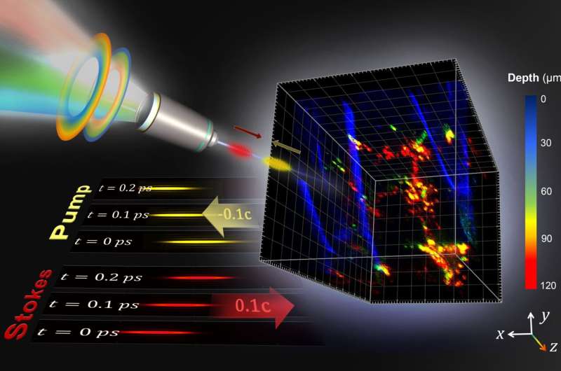
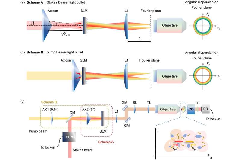
They conceived the unique angular dispersion control schemes that simultaneously convert both the pump and Stokes beam pulses into ultraslow Bessel light bullets (group velocity (vg) ~0.1c) that are independently tunable in vg using a single spatial light modulator.
More remarkably, they make the ultraslow pump and Stokes Bessel light bullets counter-propagating along the axial direction (i.e., pump: vg,pg,s>0) in the sample; thus, the depth-resolved SRS signal can be immediately detected by controlling the depth whereby the two Bessel light bullets meet with each other inside the sample through manipulating their relative time-of-flight without a need for mechanical z-scanning.
The reported technique will have broad applications for label-free deep tissue 3D chemical imaging in biological and biomedical systems and beyond.

"We proposed a novel angular dispersion control scheme using axicons and a SLM that is circularly symmetric modulated along the optical axis, thus allowing for multicolor collinear Bessel light bullets generation without aberrations for high-resolution bioimaging."
"The unique B2-SRS technique measures the interaction between the two light pulses with different vg via nonlinear optical processes, the depth information whereby the nonlinear interaction takes place can be controlled by their relative speed or relative time-of-flight for detection simply by using a regular photodetector.
"If the group velocity is vg~0.1c and pulse width ~100 fs, the resolution of the time-of-flight-based techniques should be improved by at least three orders of magnitude, up to micrometer in length, as compared to the mm-scale resolution of traditional light detection and ranging (LiDAR)," the researchers added.
"The optical sectioning method with ultraslow counter-propagating Bessel light bullet generation together with a flight-of-time resolved detection invented in B2-SRS is universal and can be readily applied to many other nonlinear optical imaging modalities to significantly advance 3D microscopy imaging in biological and biomedical systems and beyond.
"Our technique also provides new insights into the characterization of various dynamic space-time wave packets in 4D (e.g., including both spatial and temporal) with unprecedented spatiotemporal resolution and spectral information," the scientists say.
By introducing positive and negative angular dispersions on the Fourier plane, forward- and backward-propagating Stokes and pump Bessel light bullets are generated. The generated Bessel light bullets have much slower group velocities and much shorter beam lengths than conventional Bessel beam pulses. Thus, the depth-encoded SRS signal can be immediately retrieved by decoding the spatial information whereby the two Bessel light bullets meet in the sample for SRS 3D imaging.

To accomplish the task, they developed angular dispersion control schemes to generate counter-propagating Stokes and pump Bessel light bullets with positive and negative, respectively. The phase pattern displayed on the SLM is made up of two annular gratings with different grating periods and opposite blazed angles for the Stokes and pump beams.
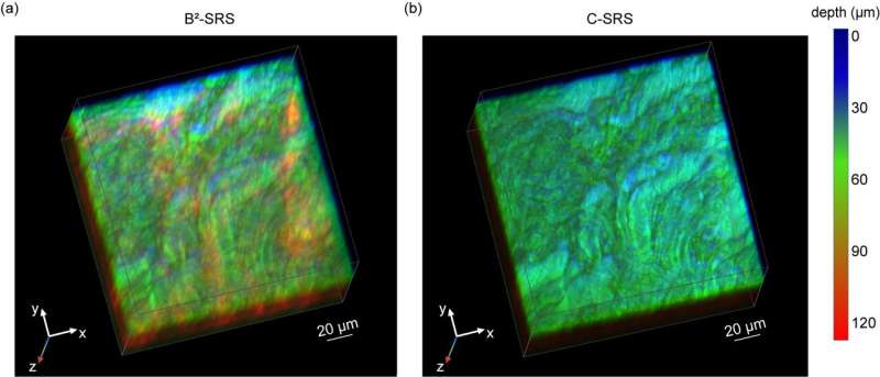
The researchers have demonstrated the advantages of the B2-SRS technique for deeper tissue 3D chemical imaging on fresh porcine brain tissue. The Bessel light bullet generated in B2-SRS has shown a remarkable scattering resilience feature in turbid media, B2-SRS gives a > 2-fold improvement in penetration depth in brain imaging compared to conventional SRS imaging.
More information: Shulang Lin et al, Time-of-flight resolved stimulated Raman scattering microscopy using counter-propagating ultraslow Bessel light bullets generation, Light: Science & Applications (2024). DOI: 10.1038/s41377-024-01498-y
Journal information: Light: Science & Applications
Provided by TranSpread


