This article has been reviewed according to Science X's editorial process and policies. Editors have highlighted the following attributes while ensuring the content's credibility:
fact-checked
peer-reviewed publication
proofread
A new and simple method for super-resolution microscopy

MINFLUX is a powerful microscopy technique that allows researchers to see objects much smaller than the wavelength of light. A newly developed evolution of the process uses a simpler device to create the light pattern needed to examine the molecule, making the entire process faster, cheaper and easier to use for future discoveries.
The research is published in the journal Light: Science & Applications.
MINFLUX pushes the boundaries of what we can see. It works by shining a specially patterned beam of light on a single molecule and measuring the intensity of the light at different locations. By analyzing these measurements, scientists can then calculate the exact position of the molecule. This allows them to study the behavior of molecules in incredible detail, providing insights into fundamental biological processes.
Unfortunately, current MINFLUX setups are complex, expensive, and require specialized equipment, which has limited the widespread use of this powerful technique. Traditional methods often involve bulky and expensive components, making MINFLUX inaccessible to many research labs.
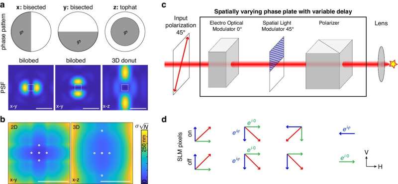
Researchers have developed a new way of creating the patterned light beam for MINFLUX. This method combines two simpler devices: a spatial light modulator (SLM) and an electro-optical modulator (EOM). The SLM acts like a digital projector, manipulating light patterns, while the EOM controls the intensity of the light. This setup is significantly faster, cheaper, and easier to use than traditional methods.
The new approach offers several advantages. Firstly, using simpler components allows for much faster scanning of the light pattern. This rapid scanning improves the accuracy of measurements, leading to sharper and more detailed images of the molecules.
Secondly, the more straightforward setup significantly reduces the cost of the equipment needed for MINFLUX, making this powerful technique more accessible to researchers. Finally, the new method is more user-friendly and can be more easily integrated into existing microscopes, streamlining the research process.
This new development paves the way for creating more affordable and accessible MINFLUX microscopes. This could open up new possibilities for studying a wide range of biological processes at the molecular level. With MINFLUX becoming more accessible, scientists can delve deeper into the unseen world, unlocking new knowledge about how life works at its most fundamental level.
More information: Takahiro Deguchi et al, Simple and robust 3D MINFLUX excitation with a variable phase plate, Light: Science & Applications (2024). DOI: 10.1038/s41377-024-01487-1
Journal information: Light: Science & Applications
Provided by TranSpread


