January 9, 2024 feature
This article has been reviewed according to Science X's editorial process and policies. Editors have highlighted the following attributes while ensuring the content's credibility:
fact-checked
trusted source
proofread
Study gathers new insight about the evolutionary origin of vertebrate jaws

Jaws are bone- or cartilage-based structures that hold together teeth in the mouth of most vertebrates (i.e., all animal species with a backbone or spinal column). While these crucial structures have been the focus of numerous studies, their evolutionary origin and their development over centuries and across species is yet to be fully elucidated.
With their strong and highly efficient jaws, sharks are one of the most emblematic animal species. Shark jaws are known to be partly flexible, for instance allowing the animals to bite off parts of their prey when they are too big for them to swallow, via a thrusting movement.
Researchers at University of Tsukuba, Ehime University and other institutes in Japan and Europe recently carried out a study examining a crucial component of the vertebrate jaw, known as the trigeminal nerve. Their paper, published in Zoological Letters, identifies unique patterns in the distribution of neuronal somata and expression of so-called Hmx genes in lamprey (also known as vampire fish), which were not present in sharks.
"The trigeminal nerve is a key component for understanding jaw evolution, as it plays a crucial role as a sensorimotor interface for the effective manipulation of the jaw," Motoki Tamura, Ryota Ishikawa and their colleagues wrote in their paper.
"This nerve is also found in the lamprey, an extant jawless vertebrate. The trigeminal nerve has three major branches in both the lamprey and jawed vertebrates. Although each of these branches was classically thought to be homologous between these two taxa, this homology is now in doubt."
The trigeminal nerve or fifth cranial nerve (V) is a key component of the vertebrate jaw, made up of both motor and sensory neurons. As suggested by its name, this nerve is typically made up of three distinct branches, namely the ophthalmic, maxillary and mandibular nerves.
Interestingly, lamprey, eel-like blood-sucking fish with sharp teeth but no clear jaw-like structure, also exhibit a trigeminal nerve. The presence of this nerve without a jaw suggests that this fish species evolved differently compared to sharks, which are known to have a conventional vertebrate jaw.
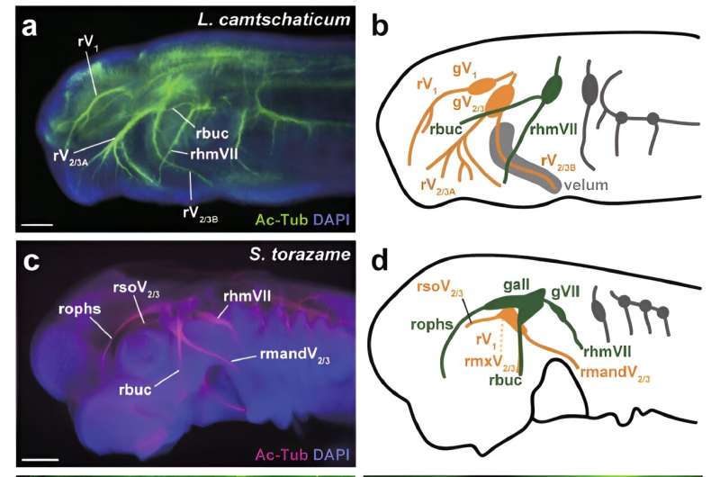
In their paper, Tamura, Ishikawa and their colleagues set out to better understand differences in the neuronal somata and gene expression patterns of lamprey and sharks. To do this, they examined the distribution of trigeminal sensory neurons in these two animals, as well as the expression patterns of Hmx genes, which are linked to the development of the nervous system and sensory organs.
Notably, previous studies on mice found that the Hmx1 gene was expressed in a specific segment of neurons, known as rV3 neuronal somata. The team tried to determine whether these same gene expression patterns were also present in sharks and lamprey.
"We compared expression patterns of Hmx, a candidate genetic marker of the mandibular nerve (rV3, the third branch of the trigeminal nerve in jawed vertebrates), and the distribution of neuronal somata of trigeminal nerve branches in the trigeminal ganglion in lamprey and shark," Tamura, Ishikawa and their colleagues wrote.
"We first confirmed the conserved expression pattern of Hmx1 in the shark rV3 neuronal somata, which are distributed in the caudal part of the trigeminal ganglion. By contrast, lamprey Hmx genes showed peculiar expression patterns, with expression in the ventrocaudal part of the trigeminal ganglion similar to Hmx1 expression in jawed vertebrates, which labeled the neuronal somata of the second branch."
Overall, the researchers found that while Hmx expression patterns and the distribution of trigeminal nerve neurons in sharks resembled those previously observed in mice, those of lamprey were markedly different. Based on these observations, they introduced two alternative hypotheses describing the possible evolutionary journey of trigeminal nerve branches.
The findings gathered as part of this study offer some new insight into the evolutionary origins of the vertebrate jaw, which remain poorly understood. In the future, they could pave the way for further works exploring the differences between the trigeminal nerve of sharks and lamprey, potentially leading to important new discoveries.
More information: Motoki Tamura et al, Comparative analysis of Hmx expression and the distribution of neuronal somata in the trigeminal ganglion in lamprey and shark: insights into the homology of the trigeminal nerve branches and the evolutionary origin of the vertebrate jaw, Zoological Letters (2023). DOI: 10.1186/s40851-023-00222-9.
© 2024 Science X Network


