This article has been reviewed according to Science X's editorial process and policies. Editors have highlighted the following attributes while ensuring the content's credibility:
fact-checked
peer-reviewed publication
trusted source
proofread
A microfluidic magnetic detection system for tumor-derived exosome analysis

In a study published on 7 November 2023 in the journal Microsystems & Nanoengineering, researchers from the Shanghai Institute of Microsystem and Information Technology, Chinese Academy of Sciences, and Shanghai Tech University, have developed an innovative microfluidic magnetic detection system (μFMS) for analyzing tumor-derived exosomes (TDEs), potential biomarkers for cancer diagnosis. This groundbreaking system could greatly enhance the early detection and treatment of cancer.
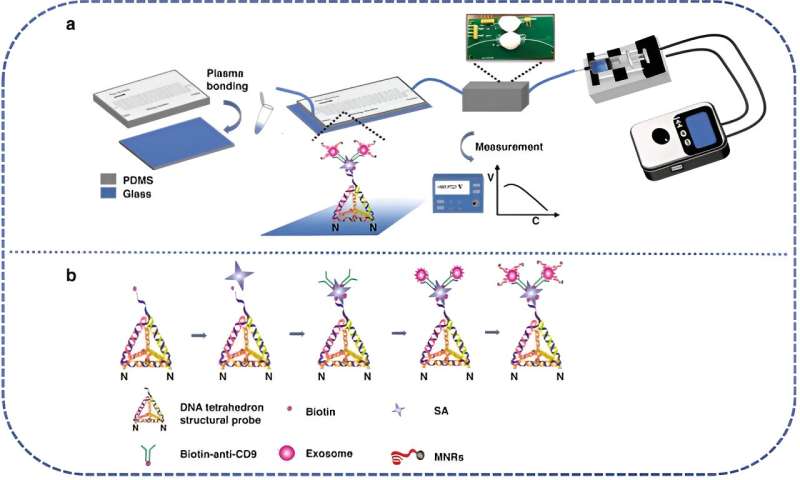
In the methodology of the microfluidic magnetic detection system (μFMS), DNA tetrahedral-structured probes (TSPs) and Fe3O4 magnetic nanoparticles (MNPs) modified with CD63 aptamers synergistically create magnetic nano-reporter probes (MNRs). These MNRs are adeptly integrated into a microfluidic chip, which features serpentine microchannels and an induction coil-based magnetic detector, thereby enabling rapid and highly sensitive detection of tumor-derived exosomes (TDEs).
The meticulous preparation process involved synthesizing specific DNA sequences to form the TSPs and MNRs. The microfluidic chips, crafted from polydimethylsiloxane (PDMS) and aldehyde-modified glass slides, are designed to immobilize TSPs effectively. Extraction of TDEs from U251 cell lines was achieved through ultracentrifugation, followed by detailed characterization using transmission electron microscopy (TEM) and dynamic light scattering (DLS).
Significant findings from this research include the μFMS's remarkable detection capabilities, exhibiting a dynamic range of 1.98 × 10³ to 1.98 × 10⁷ particles/mL and a detection limit of 1.98 × 10³ particles/mL in phosphate-buffered saline (PBS), effectively maintained even in simulated serum samples. The microfluidic chip's innovative serpentine design substantially enhances the efficiency of TDE capture, while the incorporation of DNA TSPs on the chip's surface augments the specificity of TDE detection.
The μFMS represents a major leap forward in TDE analysis. Its high sensitivity and specificity, coupled with its potential for easy integration into clinical settings, pave the way for its use in early cancer diagnosis and monitoring. The success of this system in simulated clinical serum samples reinforces its potential in real-world applications. Future research and development will focus on refining this technology for widespread clinical use, potentially transforming cancer diagnostics and therapeutics.
More information: Qiuling Qian et al, Microfluidic magnetic detection system combined with a DNA framework-mediated immune-sandwich assay for rapid and sensitive detection of tumor-derived exosomes, Microsystems & Nanoengineering (2023). DOI: 10.1038/s41378-023-00617-w
Journal information: Microsystems and Nanoengineering
Provided by Chinese Academy of Sciences


