This article has been reviewed according to Science X's editorial process and policies. Editors have highlighted the following attributes while ensuring the content's credibility:
fact-checked
peer-reviewed publication
trusted source
proofread
Scientists develop novel nanoparticles that could serve as contrast agents

Special nanoparticles could one day improve modern imaging techniques. Developed by researchers at Martin Luther University Halle-Wittenberg (MLU), the properties of these unique nanoparticles change in reaction to heat. When combined with an integrated dye, the particles may be used in photoacoustic imaging to produce high-resolution, three-dimensional internal images of the human body, the team reports in the journal Chemical Communications.
The researchers developed what are known as single-chain nanoparticles (SCNPs) which are made up of a single molecular chain and are only three to five nanometers in size. Dyes can be incorporated in these tiny capsules.
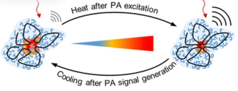
"Our SCNPs have unique thermoresponsive properties as their structure changes when exposed to heat. Depending on the temperature, the particles can take on a compact or open structure. The behavior of the encapsulated substances also changes," explains chemist Professor Wolfgang Binder from MLU, who led the study together with medical physics professor Jan Laufer, and pharmacist Karsten Mäder.
For the study, the team incorporated special dyes into the SNCPs which could then be used in photoacoustic imaging. In this type of method, laser pulses are directed at the tissue being examined. There, the energy of the light is converted into ultrasound waves, the tissue heats up, and the properties of the nanoparticles change.
When the ultrasound waves are measured outside the organism, three-dimensional images can be created that mostly show blood vessel networks. According to the researchers, the particles create a rich optical contrast that can be used, for example, to examine tumors more closely.
The team also studied how the particles functioned in cell cultures so that they could better understand if and how they work in the human body. This is crucial if the particles are to be used in biomedical applications. The novel particles performed very well in all the tests the team conducted.
"Our work is an important step in the development of thermoresponsive SCNPs, which could improve the accuracy and precision of diagnostic imaging," Binder concludes.
More information: Justus F. Thümmler et al, Thermoresponsive swelling of photoacoustic single-chain nanoparticles, Chemical Communications (2023). DOI: 10.1039/D3CC03851C
Journal information: Chemical Communications
Provided by Martin Luther University Halle-Wittenberg


