This article has been reviewed according to Science X's editorial process and policies. Editors have highlighted the following attributes while ensuring the content's credibility:
fact-checked
peer-reviewed publication
trusted source
proofread
Researchers study gut-to-CNS translocation of silver nanomaterials

Recently, a research team led by Prof. Chen Chunying from the National Center for Nanoscience and Technology (NCNST) of the Chinese Academy of Sciences (CAS) revealed that peripheral nerve fibers act as direct conduits for silver nanomaterials translocation from the gut to the central nervous systems (CNS). The study was published in Science Advances.
The transport route determines the final target organ and subsequent biological effects of exogenous nanoparticles (NPs). The specific transfer route map of NPs is crucial to unveil their nanotoxicological as well as nanomedical potential. However, there are still substantial knowledge gaps in understanding of the transport of NPs from the gut to the CNS, which hinders both the understanding of the gut-CNS axis communication and the development of new therapeutic strategies that target CNS-associated diseases.
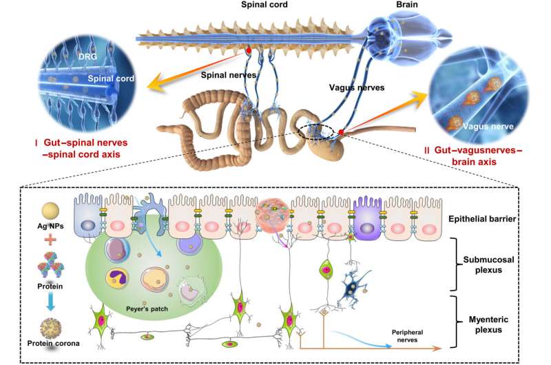
In this study, the researchers first revealed transfer routes for Ag NMs along the gut-CNS axis in addition to blood circulation. They demonstrated both theoretically and experimentally the direct translocation of Ag NMs from the gut to the CNS (both the brain and spinal cord) via peripheral nerve fibers. Using laser ablation and field flow fractionation inductively coupled plasma mass spectrometry, they showed that Ag NMs administered by oral gavage became enriched in a particle state in the brain and spinal cord of mice while not efficiently entering the blood.
In addition, the researchers found that the vagus and spinal nerves mediate the transneuronal transport of Ag NMs along the gut-brain axis and gut-spinal cord axis, respectively, with specific gut cell subsets (enterocytes and enteric nerve cells) taking up significant levels of Ag NMs for transfer to the connected peripheral nerves using Cy-TOF analysis.
The results implicated that transneuronal transport as a mechanism for the direct trafficking of NPs, filling a crucial gap in understanding the behavior of NPs in biological systems.
This study integrated a variety of methods to break through the "classical pathway" of blood circulation mediated NPs transport. For the first time, it proposed that peripheral nerve fibers as the "non-classical pathway" mediate NPs transport between organs in the body. This path of NPs in the living body provides important theoretical support for the development of nanomedicines targeting CNS-related diseases.
More information: Xiaoyu Wang et al, Peripheral nerves directly mediate the transneuronal translocation of silver nanomaterials from the gut to central nervous system, Science Advances (2023). DOI: 10.1126/sciadv.adg2252
Journal information: Science Advances
Provided by Chinese Academy of Sciences


