Fluorescence lifetime imaging for studying DNA compaction and gene activities
Fluorescence lifetime imaging for studying DNA compaction and gene activities
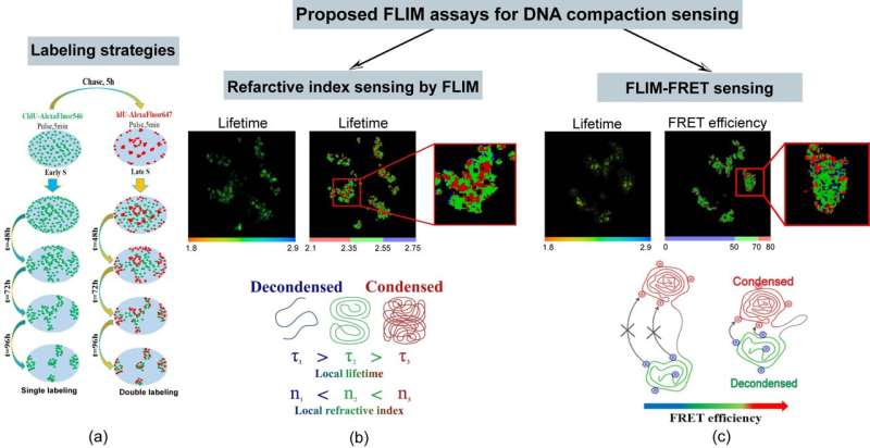
(a) Schematics of labeling approaches targeting Early S- phase and Mid S-phase replicating chromatin domains. Labeling protocol was adjusted for segregation of the labeled chromosome territories to facilitate image analysis. (b) and (c) FLIM sensing of chromatin refractive index and FLIM-FRET sensing of the DNA compaction, respectively. Credit: by Svitlana M. Levchenko, Artem Pliss, Xiao Peng, Paras N. Prasad and Junle Qu

Studies of the genomic DNA compaction in the cell nucleus and dynamic reorganization during physiologic processes or disease development in live cell environments are exceedingly challenging. This complexity stems from a high degree of compaction required to accommodate ~2 meters of genomic DNA within the cell nucleus, which typically is 5 to 10 micrometers in diameter. In addition, the chromatin compaction density is not static, but fluctuates over time accommodating for gene activities. Meanwhile, the 3D resolution of optical microscopy is not high enough even for sub-diffraction imaging modalities, thus limiting studies of the spatial geometry of complex genomic architecture and its dynamic transformations.
Fluorescence lifetime imaging for studying DNA compaction and gene activities
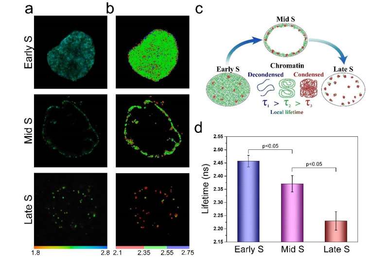
(a) Representative fluorescence lifetime images of Early-, Mid- and Late S-phase DNA replication sites labeled with AlexaFluo 546. (b) Color-coded image of the lifetime distribution throughout cell nucleus. (c) Schematics of the FLIM approach to measure compaction of chromatin replicating at different S-phase windows. (d) Averaged fluorescence lifetime of AlexaFluo546 used to label DNA replication sites in Early-, Mid-, and Late S-phase. The error bars correspond to standard deviations. Data indicate the higher refractive index, and thus higher compaction density for gene-poor chromatin domains replicating in Late S-phase compared to gene-rich chromatin replicated in Early S-phase. Credit: Svitlana M. Levchenko, Artem Pliss, Xiao Peng, Paras N. Prasad and Junle Qu

In a new paper published in Light: Science & Applications, a team of scientists, led by Professor Junle Qu from Center for Biomedical Optics and Photonics & College of Physics and Optoelectronic Engineering, Shenzhen University, China, and Prof. Paras N. Prasad from Institute of Lasers, Photonics and Biophotonics, State University of New York at Buffalo, U.S., has developed an alternative strategy that is based on fluorescence lifetime imaging (FLIM) to overcome existing limitations of conventional approaches. The authors propose two independent FLIM assays enabling fine measurements of DNA compaction. The first one relies on the inverse quadratic relation between the fluorescence lifetimes of fluorescent probes incorporated into DNA and their local refractive index, variable due to chromatin compaction density. Another FLIM approach employs Förster resonance energy transfer (FRET) between the fluorescence labeled nucleotides incorporated into the DNA strands.
Fluorescence lifetime imaging for studying DNA compaction and gene activities
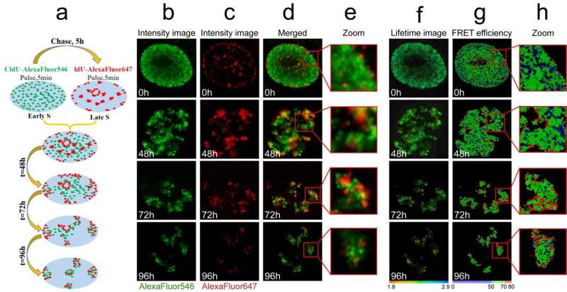
(a) Schematics of approach: cultured cells sequentially labeled with CldU in early S-phase and with IdU in the late S-phase were followed into subsequent cell generations, enabling to visualize segregated chromosome territories with stained early and late S-phase labeled chromatin. Label segregation enables analysis of chromatin compaction within individual chromosome territories. (b-c) Cells were fixed at various intervals following the exposure to halogenated nucleotides and stained (b) for CldU (AlexaFluor 546, green) and (c) IdU (AlexaFluor647, red) as shown in the panel (a). Representative fluorescence intensity (b-e), lifetime images (f) and FRET efficiency (g-h) of the chromosome territories formation process after labeling of the Early and Late S-phase replicating chromatin domains. High FRET areas (red) indicate a close proximity between the chromatin fibers replicated in Early- and Late S-phase. Credit: Svitlana M. Levchenko, Artem Pliss, Xiao Peng, Paras N. Prasad and Junle Qu

In this study, both FLIM assays were validated in cultured cells, where the researchers comparatively analyzed compaction of gene-rich chromatin domains that replicate in Early S-phase, and those that replicate in Mid- to Late S-phase and contain predominantly non-coding sequences. The obtained data demonstrate the sensitivity of both FLIM assays and unravel a significant difference in compaction of the gene-rich and gene-poor pools of genomic DNA. They show that the gene-rich DNA is loosely compacted compared to the dense DNA domains devoid of active genes.

More information: Svitlana M. Levchenko et al, Fluorescence lifetime imaging for studying DNA compaction and gene activities, Light: Science & Applications (2021). DOI: 10.1038/s41377-021-00664-w

Journal information: Light: Science & Applications

Citation: Fluorescence lifetime imaging for studying DNA compaction and gene activities (2021, December 27) retrieved 17 July 2024 from https://phys.org/news/2021-12-fluorescence-lifetime-imaging-dna-compaction.html
This document is subject to copyright. Apart from any fair dealing for the purpose of private study or research, no part may be reproduced without the written permission. The content is provided for information purposes only.

Explore further

Fluorescence lifetime imaging to study DNA compaction and gene activities

18 shares

Feedback to editors