The mechanics of cavitation-induced injury lend a better understanding of blast traumatic brain injuries

Traumatic brain injury (TBI) is a largely silent epidemic that affects roughly two million people each year, according to the U.S. Centers for Disease Control and Prevention. But the scale at which blast TBI (bTBI) injuries—in the spotlight as the signature wound of the wars in Iraq and Afghanistan—occur and manifest is unknown.
Recent studies within this realm suggest that rapid cavitation bubble collapse may be a potential mechanism for studying bTBI.
During the Biophysical Society's 61st Annual Meeting, Feb. 11-15, 2017, in New Orleans, Louisiana, Jonathan Estrada, a doctoral student in the School of Engineering at Brown University, will present his work exploring the mechanics of cavitation-induced injury—with a goal of better understanding bTBIs.
Estrada is working under the guidance of Christian Franck, along with colleagues from Brown University and the University of Michigan. The team uses a laser, an optical microscope and rat neurons inside a gel-like substance to mimic brain tissue to examine bTBIs.
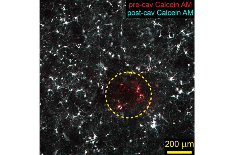
The laser pulse is sent through the "brain tissue" under the microscope while a high-speed camera—recording 270,000 frames per second—captures the laser creating a bubble, the bubble breaking and the damage this causes to the rat neurons. "We image affected neurons before and immediately after injury," Estrada said.
The significance of the group's work is that while postmortem studies have begun to show differences in brain pathology—such as astroglial (star-shaped glial cell) scarring—between patients exposed to blast injury and those with blunt TBI, the manifestation of injury over time still isn't well understood. "Our work, using the simplified bubble and neuron culture model, aims to begin bridging the gap between the mechanics of blast injury and cell damage," Estrada said.
Although the results are in the preliminary stage, "so far, we've found that the maximum bubble radius is nearly identical to the zone of neuron fragmentation immediately after injury," he added. "This is in contrast to a previous study from our group that focused on concussive, or blunt, TBI via uniaxial compression of neurons, which found that injury was distributed over entire cultures rather than localized to one area."
In terms of applications, the group's method allows them to see the injury history of the cells within cultures—before and just after injury with live-cell fluorescence, during injury with high-speed imaging, and then injury manifestation at later time points via immunostaining. "Quantifying temporal injury history is essential to understanding, diagnosing, and working toward informed treatment of blast TBI," Estrada noted. "We hope this is a positive step toward those ends."
More information: Microcavitation as a neuronal damage mechanism in an in vitro model of blast traumatic brain injury. www.abstractsonline.com/pp8/#! … 79/presentation/1225
Provided by Biophysical Society


