Neuroscientists decode the brain activity of the worm

Manuel Zimmer and his team at the Research Institute of Molecular Pathology (IMP) present new findings on the brain activity of the roundworm Caenorhabditis elegans. The scientists were able to show that brain cells (neurons), organized in a brain-wide network, albeit exerting different functions, coordinate with each other in a collective manner. They could also directly link these coordinated activities in the worm's brain to the processes that generate behavior. The results of the study are presented in the current issue of the journal Cell.
One of the major goals of neuroscience is to unravel how the brain functions in its entirety and how it generates behavior. The biggest challenge in solving this puzzle is represented by the sheer complexity of nervous systems. A mouse brain, for example, consists of millions of neurons linked to each other in a highly complex manner. In contrast to that, the nematode Caenorhabditis elegans is equipped with a nervous system comprised of only 302 neurons. Due to its easy handling and its developmental properties, this tiny, transparent worm has become one of the most important model organisms for basic research. For almost 30 years, the list of connections between individual neurons has been known. Despite the low number of neurons, its neuronal networks possesse a high degree of complexity and sophisticated behavioral output; the worm thus represents an animal of choice to study brain function.
Interplay of neuronal groups in brain-wide networks
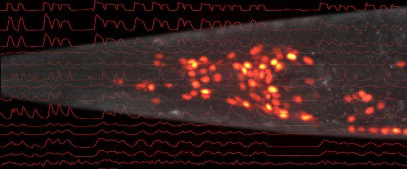
Researchers have mostly concentrated on studying the functions of single or a handful of neural cells and some of their interactions to explain behavior such as movements. For the worm, it has been known how some single neurons function as isolated units within the network, but it remained unknown how they work together as a group. Manuel Zimmer, a group leader at the IMP, wanted to address this unsolved question in his research. Together with his team, he combined two state-of-the-art technologies for the current study: first, the scientists used 3D microscopy techniques to simultaneously and rapidly measure different regions of the brain; second, they used worms genetically engineered with a fluorescent protein that caused the worm's neurons to flash when they were active. "This combination was brilliant for us, as it allowed a brain-wide single-cell resolution of our recordings in real time," Zimmer explains the advantages of this approach.
Reading the worm's mind
Zimmer and his team tested the animals' reaction to stimuli from outside when they were trying to find food. Under the microscope, a fascinating picture was revealed to the researchers: "We saw that most of the neurons are constantly active and coordinate with each other in a brain-wide manner. They act as an ensemble", explains postdoctoral scientist Saul Kato, who spearheaded the study together with Harris Kaplan and Tina Schrödel, graduate students in the Zimmer laboratory. The animals were immobilized for these experiments, their reactions therefore representing intentions as opposed to reflecting actual movement.
With a different technique of microscopy, set up for freely moving worms, the scientists were able to detect the neurons that initiate movement. There was a direct correlation between the activity of certain networks and the impulse for movements; thus Zimmer and his co-workers could literally watch the worms think. These network activities not only represented short movements, but also their assembly into longer lasting behavioral strategies such as foraging. "This is something that no one has managed to do before", Zimmer points out. Suggestions of similar patterns of neural activity have been found in higher animals, but so far only a fraction of neurons in sub-regions of the brain could be examined at the same time. Zimmer and his colleagues are therefore confident that their results represent basic principles of brain function, even though the worm is only distantly related to mammals.
Investigation of molecular mechanisms
Many questions in the area of neurobiology remain largely unsolved, such as how decisions are made or whether the brain operates in a formal algorithmic manner, like a computer. In the next phase of research, Manuel Zimmer intends to analyze the molecular mechanisms underlying the processes he investigated. "It would also be interesting to have a closer look at long lasting brain states such as sleep and waking", he says, laying out his ambitious plans for the future.
More information: Kato et al., Global Brain Dynamics Embed the Motor Command Sequence of Caenorhabditis elegans, Cell (2015), dx.doi.org/10.1016/j.cell.2015.09.034
Journal information: Cell
Provided by Research Institute of Molecular Pathology


