Scientists develop atomic force microscopy for imaging nanoscale dynamics of neurons

Researchers at the Max Planck Florida Institute for Neuroscience and Kanazawa University (Japan) have succeeded in imaging structural dynamics of living neurons with an unprecedented spatial resolution.
While progress has been made over the past decades in the pursuit to optimize atomic force microscopy (AFM) for imaging living cells, there were still a number of limitations and technological issues that needed to be addressed before fundamental questions in cell biology could be address in living cells.
In their March publication in Scientific Reports, researchers at Max Planck Florida Institute for Neuroscience and Kanazawa University describe how they have built the new AFM system optimized for live-cell imaging. The system differs in many ways from a conventional AFM: it uses an extremely long and sharp needle attached to a highly flexible plate. The system is also optimized for fast scanning to capture dynamic cellular events. These modifications have enabled researchers to image living cells, such as mammalian cell lines or mature hippocampal neurons, without any sign of cellular damage.
"We've now demonstrated that our new AFM can directly visualize nanometer-scale morphological changes in living cells", explained Dr. Yasuda, neuroscientist and scientific director at the Max Planck Florida Institute for Neuroscience.
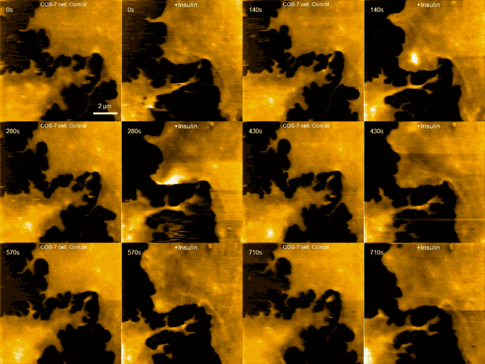
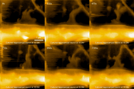
In particular, this study demonstrates the capability to track structural dynamics and remodeling of the cell surface, such as morphogenesis of filopodia, membrane ruffles, pit formation or endocytosis, in response to environmental stimulants. An example of this capability can be visualized in movie 1, where a fibroblast is imaged before and after treatment with insulin hormone, which intensely enhances the ruffling at the leading edge of the cell. Another example is seen in movie 2, where the morphological changes of a finger-like neuronal protrusion in the mature hippocampal neuron are observed.

According to Dr. Yasuda, the successful observations of structural dynamics in live neurons present the possibility of visualizing the morphology of synapses at nanometer resolution in real time in the near future. Since morphology changes of synapses underlie synaptic plasticity and our learning and memory, this will provide us with many new insights into mechanisms of how neurons store information in their morphology, how it changes synaptic strength and ultimately how it creates new memory.
More information: Scientific Reports, www.nature.com/srep/2015/15030 … /full/srep08724.html
Journal information: Scientific Reports
Provided by Max Planck Society


