Scientists develop compact medical imaging device
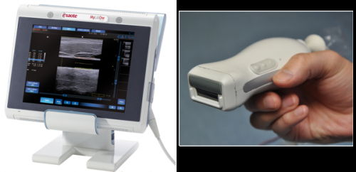
Scientists at the MIRA research institute, in collaboration with various companies, have developed a prototype of a handy device that combines echoscopy (ultrasound) with photoacoustics. Combining these two medical imaging technologies in a compact device is designed, among other things, to enable the amount of inflammation in rheumatic patients' joints to be measured more simply and precisely. The researchers expect that the technology will eventually also be able to play a role in detecting the severity of burns, skin cancer and furring of the arteries. The prototype is presented in the scientific journal Optics Express.
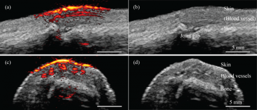
Echoscopy and photoacoustics are complementary medical imaging technologies. Photoacoustics involves sending brief laser pulses into the patient's body. When the laser light hits a blood vessel, for example, it is locally converted into heat, which causes a minor rise in pressure. This propagates through the body like a sound wave and can then be measured on the skin. Echoscopy involves sending ultrasound waves into the body: different tissues reflect them in different ways, and they too can then be detected on the skin. Whereas echoscopy provides an image of structures, photoacoustics can provide an image containing more functional information, such as the presence of blood.
Combination
Combining the two technologies in one device enables images of the body to be produced that contain far more information. The scientists have made the combination possible by integrating pulsing diode lasers in the ultrasound transducer, resulting in a compact and relatively inexpensive system. The measuring technique works best on relatively superficial parts of the body, down to a depth of fifteen millimetres.
Applications
In the article published in Optics Express the researchers show that photoacoustic and ultrasound images of tissue structure and function can be produced in real time. They therefore expect the device, which has taken them about four years to develop, to be able to measure the amount of inflammation in rheumatic patients' joints more precisely. The MIRA researchers will be looking into this in the coming years in collaboration with the ZGT hospital group. They will also be investigating in a European context whether the technique could also be used to ascertain the severity of burns and their responsiveness to treatment, and to detect skin cancer and furring of the arteries. The next step is to develop a prototype that enables deeper measurements to be carried out and more functional information to be obtained, such as blood oxygen saturation.
More information: "Handheld probe integrating laser diode and ultrasound transducer array for ultrasound/photoacoustic dual modality imaging." Optics Express, Vol. 22, Issue 21, pp. 26365-26374 (2014) dx.doi.org/10.1364/OE.22.026365
Journal information: Optics Express
Provided by University of Twente


