New imaging technique leads to better understanding of freezing in plants

Using a new technique to study an old problem, an Agricultural Research Service scientist in North Carolina has uncovered new details about what happens to a cereal plant when it freezes.
Agronomist David P. Livingston, in the Plant Science Research Unit in Raleigh, has developed an imaging technique and has used it to show that when an oat plant freezes, ice forms in its roots and in portions of its crown, which lies just below the soil surface and connects the roots to the stalk.
The results have implications for growers. In winter cereals like oats (Avena sativa), the crown is where the plant generates new tissue growth—if it survives the winter cold. Oats won't grow in many northern areas because of cold temperatures. Understanding how ice forms in oats could help breeders develop hardier varieties and expand their range, Livingston says. Climate change has also made it more important to understand how cereal crops react to wide fluctuations of winter temperatures and other environmental stresses.
The process developed by Livingston involves making high-resolution digital photos of standard histological slices of plant tissues and using commercially available software to create a three-dimensional perspective, which gives added depth to their structures, above and below ground. The resulting images are similar to those produced by magnetic resonance imaging (MRI) and computed tomography (CT) scans. The advantages of Livingston's images are that they can be created from much smaller tissue samples than what CT and MRI tests require and are less expensive to produce, because they require less expensive equipment and training.
Livingston's studies have so far focused on oats because their production in the United States is limited by their sensitivity to subfreezing temperatures. He has also used the technique to examine wheat, barley, rye, and corn, and he says it could be used to study other crop plants. The technique even works on mammalian systems and has been used to produce three-dimensional reconstructions of tumors in liver biopsies.
 as seen in this interior view. Credit: David Livingston.")
Livingston first described the technique in a paper in the Journal of Microscopy in 2010, which included images of oat tissue and lung tissue from a mouse. In the more recent study, he used it to examine how oat plants react to freezing temperatures in the soil. He stained frozen tissue samples and took 186 sequential images of them with a digital camera. He then aligned the images and used imaging software to clear away the background colors so he could focus on cavities formed by ice crystals in the crown tissues of the oats. He compared the images from frozen plants with images from plants kept at normal temperatures.
Along with showing how ice forms in the root, the images revealed that ice formation in the crown is limited to its lowest and uppermost parts, apparently leaving the middle free of ice—at least free from crystals big enough to visualize. The ice also didn't form in the shape of circular crystals, as portrayed in two-dimensional images. Instead, the crystals were shaped more like elongated curtains.
The results were published in 2014 in Environmental and Experimental Botany.
-
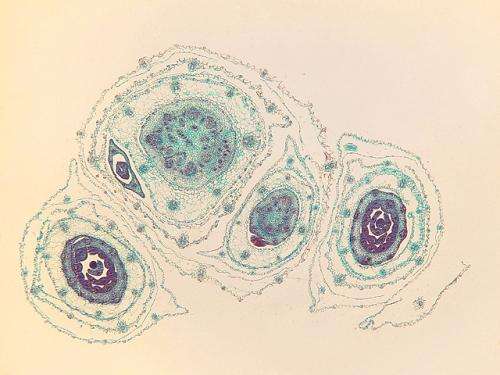
Cross-section views of a plant crown before freezing. Credit: David Livingston -
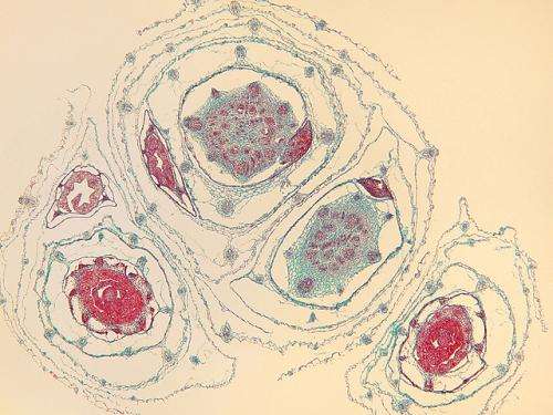
After the plant crown was frozen and thawed, some empty spaces that were not present before freezing exist within the tissue where ice had formed while the plant tissue was frozen. Credit: David Livingston -
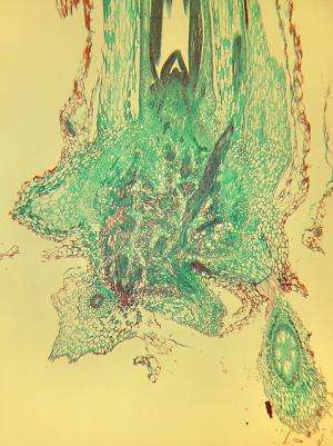
A longitudinal section of the crown area showing the complexity of tissue within it. Credit: David Livingston
Provided by Agricultural Research Service


