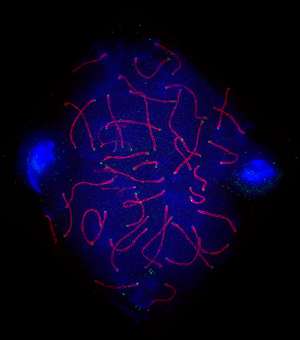
Prize-winning microscopy image lights up Times Square in New York

Science and show business may sound like an unusual combination, but advances in technology mean that scientists can now capture dramatic images of their research that easily match the glamor of Broadway. A striking image captured by Graham Wright and Henning Horn from the A*STAR Institute of Medical Biology (IMB) in Singapore during their ground-breaking investigations into fertility was the regional winner in the microscopy category of the 2013 GE Healthcare Life Sciences Cell Imaging Competition. Fittingly, together with the other prize-winning images, the image was recently displayed on a large, high-resolution screen in New York's iconic Times Square.
"The image is of a mouse sperm cell, also known as a spermatocyte, highlighted with three fluorescent labels that show DNA (blue), KASH5 protein (green) and the SCP3 protein (red), which is required for the pairing of chromosomes," explains Wright. "This image was a particularly striking example when we captured it—the orientation of the proteins we were studying and the two sperm cells stained in blue on both sides made it aesthetically pleasing."
The image was the result of a collaboration between Wright, head of the IMB Microscopy Unit (IMU), Horn, a senior research fellow, and the research teams of Colin Stewart and Brian Burke, also of the IMB, who discovered that the KASH5 protein is vital for successful chromosomal movements during meiosis—the division of cells necessary for successful sexual reproduction1. Sperm and eggs need accurate chromosome pairing if they are to mature correctly, so without chromosomal activity guided by the KASH5 protein, fertility is adversely affected.
The researchers collected the image on a GE DeltaVision OMX microscope, which enables biological samples to be imaged in superresolution in three dimensions. Wright and Horn spent time perfecting their sample preparation and honing the settings on the microscope to acquire their high-resolution prize-winning image. "The increased resolution we were able to achieve allowed us to visualize chromosome pairing events in spermatocytes."
"Paired chromosomes—the paired red lines in the image—are not resolved by conventional light microscopy techniques," explains Horn. "The ability to determine whether chromosomes are paired or not was critical for understanding the function of KASH5 in meiosis, so images such as this really can change how we understand diseases and problems such as infertility."
At the IMB, research is focused on understanding a number of human diseases and medical conditions and providing improved treatments. Researchers have direct access to state-of-the-art equipment, including high-end microscopes, through core technology platforms such as the IMU.
By gaining further insight into the processes behind meiosis using these advanced microscopy techniques, Wright and Horn hope that their work will shed light on a range of biological processes, including those underpinning human fertility problems.
"Microscopy is usually used to visualize and analyze how proteins, cells and tissues are organized and how they behave," states Wright. "But with fluorescent dyes and live-cell imaging techniques, we can find where proteins are located and follow what they do over time. This can give us excellent insights into the function of a protein and what goes wrong with it in the diseases we study."
More information: Horn, H. F., Kim, D. I., Wright, G. D., Wong, E. S. M., Stewart, C. L. et al. "A mammalian KASH domain protein coupling meiotic chromosomes to the cytoskeleton." The Journal of Cell Biology 202, 1023–1039 (2013). dx.doi.org/10.1083/jcb.201304004
Journal information: Journal of Cell Biology


