New method to analyse how cancer cells die
(Phys.org) —A team from The University of Manchester – part of the Manchester Cancer Research Centre - have found a new method to more efficiently manufacture a chemical used to monitor cancer cells.
The technique could lead to clearer and better quality images on PET scans.
The number of cells within tissue is controlled through apoptosis – a process where cells shrink and their components break up, also known as programmed cell death. Cancer is often characterised by a disruption to the normal process of this cell death.
Being able to study this process accurately would allow doctors to more effectively diagnose and monitor cancer and to test and develop new treatments designed to kill cancer cells.
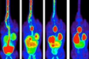
Ideally, cell death would be measured non-invasively to avoid surgery and current methods are focused on using radioactive tracers – molecules that are taken up in regions of tissue where cells are breaking apart as they die. These tracers release radiation that can be detected using a scanner. The resulting scan images show where the tracer is concentrated and therefore where cell death activity is highest.
One such tracer – ML-10 – accumulates where there are dying cells. By labelling this molecule with radioactive fluorine – that results in gamma rays that can be detected by scanners - and injecting it into patients, scientists can detect where cell death is taking place using a PET scan.
Manchester researchers have developed a new manufacturing process for ML-10, and tested the tracer in mice, and have also shown how to make an alternative, potentially better, tracer with a similar chemical structure.
Dr Gavin Brown, who led the research, said: "Measurement of cell death could help doctors monitor new anticancer therapies. We have devised a new way to manufacture this tracer, and an alternate form, that could potentially improve the quality of PET images."
The team reported their results recently in the journal Bioorganic and Medicinal Chemistry. Their method involves several stages of carefully reacting chemicals together in order to achieve the required product.
They found that their new method was better at making the tracer stick to dying cells – this will allow medics to monitor what is happening and makes it easer to detect the dying cells.
In addition, they showed that the alternate form of ML-10 could also be used to detect cell death.
"This new chemical will lead to clearer and better quality images. The preliminary results we have achieved are very promising and we feel that our new approach warrants further investigation," added Dr Brown.
More information: Kadirvel M, et al. "Detection of apoptosis by PET/CT with the diethyl ester of [¹⁸F]ML-10 and fluorescence imaging with a dansyl analogue." Bioorg Med Chem. 2014 Jan 1;22(1):341-9. DOI: 10.1016/j.bmc.2013.11.019 . Epub 2013 Nov 20.
Provided by University of Manchester


