Nano-capsules designed for diagnosing malignant tumours

Japanese researchers have developed adaptable nano-capsules that can help in the diagnosis of glioblastoma cells – a highly invasive form of brain tumour.
Polymersomes are hollow, synthetic, nano-sized capsules. They have been extensively studied for their potential in the targeted delivery of drugs within the body. PICsomes are a novel class of polymersomes that were recently developed in Japan. They are made by mixing electrolyte groups formed of positively and negatively charged ions. PICsomes can survive in the bloodstream for long periods and can be used to deliver water-soluble substances to target tissues.
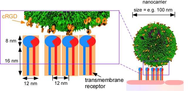
A study published in Science and Technology of Advanced Materials (STAM) reports that PICsomes can be adapted to last longer in the bloodstream and to better target specific sites in tumours. These "functionalized" PICsomes have great potential for use in both drug delivery and in the magnetic resonance imaging of tumours.
Cyclic RGD (cRGD) – a peptide or short chain of amino acids – is known to specifically bind to two receptors that play a significant role in the formation of new blood vessels in tumours. This makes it a good tumour tracer. In the STAM study, Japanese researchers bound cRGD to PICsomes. The cRGD-PICsomes were then intravenously injected into mice that had been inoculated – under the skin – with human glioblastoma cells, a highly invasive brain tumour. The research team found that the cRGD-PICsomes accumulated mainly in the new blood vessels of the tumour and that they remained near these blood vessels 24 hours later.
The researchers then loaded cRGD-PICsomes with superparamagnetic iron oxide (SPIO), which is used to improve the visibility of internal body structures in magnetic resonance imaging. In addition, glioblastoma cells were injected into the brains of mice and allowed to grow for more than two weeks. The SPIO-loaded cRGD-PICsomes were then intravenously injected into the mice. Using magnetic imaging, the team successfully tracked the loaded PICsomes in new blood vessels that formed around the glioblastomas.
Previous research has shown that magnetic imaging can detect SPIO-loaded PICsomes that are not bound with cRGD in vascular-rich tumours such as tumours of the colon. But they can't always be detected in glioblastomas: brain tumours that are strongly protected by the blood-brain barrier, which prevents both toxic substances and drugs from reaching the brain.
The results of the study suggest that SPIO-loaded cRGD-PICsomes might be useful for enhancing the contrast during magnetic resonance imaging in tumour microenvironments, including in new blood vessels that overexpress cRGD-sensitive transmembrane receptors.
Because blood vessel formation is intimately related to tumour malignancy, magnetic resonance imaging using PICsomes that are adapted to target tumour blood vessels could be a promising tool for the accurate diagnosis of malignant tumours.
More information: "Density-tunable conjugation of cyclic RGD ligands with polyion complex vesicles for the neovascular imaging of orthotopic glioblastomas." Science and Technology of Advanced Materials DOI: 10.1088/1468-6996/16/3/035004
Journal information: Science and Technology of Advanced Materials
Provided by National Institute for Materials Science


