December 12, 2014 report
Researchers use gold substrate to allow for electron cryomicroscopy on difficult proteins

(Phys.org)—A pair of researchers with the Medical Research Council Laboratory of Molecular Biology at the University of Cambridge in the U.K. has found a way to overcome the problem of movement by many proteins when attempting to study them using electron cryomicroscopy. In their paper published in the journal Science, Christopher Russo and Lori Passmore describe how they created a support structure based on a gold substrate that appears to solve the problem.
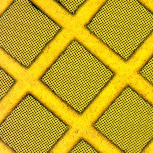
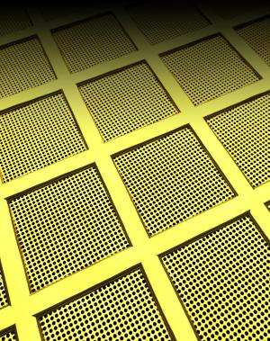
As the research pair note, up till now it's been difficult to learn more about many proteins using electron cryomicroscopy because they move around under the beam, which results in blurry images and difficulty in making measurements. They further explain that the reason this occurs is because of instabilities in the carbon substrates generally used to support samples under the microscope (which are frozen to increase stability.) In this new effort, the researchers describe their new technique, which they believe offers a way around this problem by introducing support substrates made from gold. They fashioned a support structure that was roughly similar to those based on carbon substrates then stretched a gold film just 500 angstroms thick over a square lattice support—they finished by placing the frozen proteins into 1.2 micrometer sized holes they'd made in the film. The film and gold support, they noted, ensured uniform electrical conductivity and thermal contraction as the protein samples were frozen.
The research pair suggest that gold lends itself to the application for several reasons—it's highly resistant to radiation and thus more stable, it's chemically inert and is both biocompatible and electrically conductive.
To test their process, they scanned apoferritin—a protein with a spherical complex that has proven exceptionally difficult to study with electron cryomicroscopy, using both the old technique and the new one they'd developed. They report that during the test with the gold substrate, the sample exhibited very little motion, (both in the vertical and object plane) allowing for much better images to be taken than they were able to get when they used a carbon substrate. They also report that because of the reduced motion, they were able to move closer to the sample as the scans were being made. They believe their method will likely work with a whole host of other proteins which should keep other researchers busy for quite a long time.

More information: Ultrastable gold substrates for electron cryomicroscopy, Science 12 December 2014: Vol. 346 no. 6215 pp. 1377-1380, DOI: 10.1126/science.1259530
ABSTRACT
Despite recent advances, the structures of many proteins cannot be determined by electron cryomicroscopy because the individual proteins move during irradiation. This blurs the images so that they cannot be aligned with each other to calculate a three-dimensional density. Much of this movement stems from instabilities in the carbon substrates used to support frozen samples in the microscope. Here we demonstrate a gold specimen support that nearly eliminates substrate motion during irradiation. This increases the subnanometer image contrast such that α helices of individual proteins are resolved. With this improvement, we determine the structure of apoferritin, a smooth octahedral shell of α-helical subunits that is particularly difficult to solve by electron microscopy. This advance in substrate design will enable the solution of currently intractable protein structures.
Journal information: Science
© 2014 Phys.org


