Related topics: cells

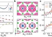
Physicists reach atomic-scale telegraphy with light

In the 1880s Heinrich Hertz discovered that a spark jumping between two pieces of metal emits a flash of light—rapidly oscillating electromagnetic waves—which can be picked up by an antenna. To honor his groundbreaking ...

Seeing is believing: Observation of migrasomes

Migrasomes, novel organelles first reported by Professor Li Yu' s team in 2015, are vesicular structures with diameters ranging from 0.5 to 3 micrometers that form on the retraction fibers at the rear of migrating cells. ...

Microscopy

Microscopy is the technical field of using microscopes to view samples or objects. There are three well-known branches of microscopy, optical, electron and scanning probe microscopy.

Optical and electron microscopy involve the diffraction, reflection, or refraction of electromagnetic radiation/electron beam interacting with the subject of study, and the subsequent collection of this scattered radiation in order to build up an image. This process may be carried out by wide-field irradiation of the sample (for example standard light microscopy and transmission electron microscopy) or by scanning of a fine beam over the sample (for example confocal laser scanning microscopy and scanning electron microscopy). Scanning probe microscopy involves the interaction of a scanning probe with the surface or object of interest. The development of microscopy revolutionized biology and remains an essential tool in that science, along with many others including materials science and numerous engineering disciplines.

This text uses material from Wikipedia, licensed under CC BY-SA