This article has been reviewed according to Science X's editorial process and policies. Editors have highlighted the following attributes while ensuring the content's credibility:
fact-checked
peer-reviewed publication
trusted source
proofread
Review covers optical aspects of quantitative photoacoustic tomography

Quantitative photoacoustic tomography (QPAT) is a medical imaging technique that combines laser-induced photoacoustic signals and ultrasound detection to create detailed three-dimensional images of biological tissues. The process involves irradiating biological tissues with short laser pulses. These pulses are absorbed by light-absorbing molecules (chromophores) within the tissues, leading to rapid heating and the generation of ultrasonic waves or acoustic signals.
The resulting distribution of acoustic pressure is measured and recorded over time, forming a photoacoustic time series used for reconstructing a three-dimensional tissue image. In photoacoustic tomography, laser pulses are dispersed over a wider tissue area rather than being focused on a specific region. To produce the final tissue image, it is crucial to estimate the optical properties of tissues from the measured photoacoustic time-series.
In a review published in the Journal of Biomedical Optics (JBO), Tanja Tarvainen from the University of Eastern Finland and Ben Cox from University College London discuss the optical part or image generation aspect of QPAT.
"Our study is focused on the mathematics of the optical part," says Tarvainen. "It surveys the current thinking regarding two related problems: what is the best way to describe light propagation and its interaction with biological tissue mathematically? Given photoacoustic measurements, what, in principle, can we learn about the optical properties of tissue, or indeed the related and more clinically relevant properties such as blood oxygenation?"
The review begins by introducing commonly used mathematical models for describing the propagation of light and sound in biological tissues, specifically the radiative transfer equation (RTE) and its approximations. These equations describe the movement of light through a medium, considering its absorption, scattering, and emission. In QPAT, the RTE serves as a model to understand how light interacts with biological tissues, assuming the constant energy of photons during elastic collisions and a constant refractive index of the medium.
The review then introduces the Grüneisen parameter, which links the optical energy absorbed by the tissues to the initial acoustic pressure distribution. Equations for propagation of the acoustic waves in biological tissue are highlighted as well.
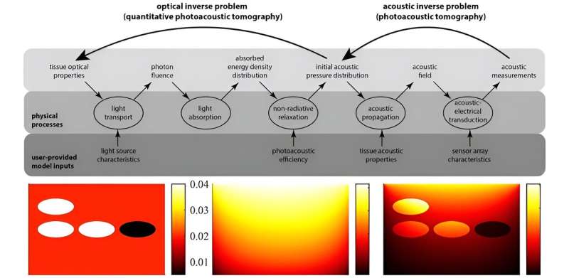
Next, the researchers discuss the photoacoustic inverse problem which involves estimating the concentrations of light-absorbing molecules in biological tissues. There are two inverse problems in QPAT. In the acoustic inverse problem, the acoustic pressure distribution is determined from the measured photoacoustic time series.
However, this review focuses on the optical inverse problem, where the distributions of optical parameters are estimated from the absorbed optical energy density. Solving inverse problems is important for obtaining accurate estimates of clinically important parameters, such as the concentrations of oxyhemoglobin and deoxyhemoglobin, which are indicators of blood oxygen saturation levels.
The authors outline two approaches to the optical inverse problem in QPAT: a direct estimation of chromophore concentrations from absorbed optical energy density data and a two-stage process involving the recovery of absorption coefficients, followed by spectroscopic inversion to calculate the concentration.
Finally, the review discusses the challenges associated with the practical implementation of QPAT. These include addressing the effect of optical scattering, considering the variation in the absorption of optical energy by the tissues (fluence effect), the need for intensive computational methods, and uncertainties in parameters that are used as inputs to the models, such as the Grüneisen parameter.
"Although QPAT is a promising methodology for providing high-resolution 3D images of physiologically relevant parameters, there are many computational modeling-based challenges that need to be tackled before the technique can be developed as a standard clinical or preclinical tool," says Tarvainen.
QPAT holds significant promise for non-invasive medical imaging and diagnosis. The topics discussed in the present review can guide the development of strategies to enhance the accuracy and reliability of QPAT in real-world scenarios.
More information: Tanja Tarvainen et al, Quantitative photoacoustic tomography: modeling and inverse problems, Journal of Biomedical Optics (2023). DOI: 10.1117/1.JBO.29.S1.S11509
Journal information: Journal of Biomedical Optics
Provided by SPIE


