This article has been reviewed according to Science X's editorial process and policies. Editors have highlighted the following attributes while ensuring the content's credibility:
fact-checked
peer-reviewed publication
trusted source
proofread
Rolling Fourier ring correlation method maps local quality at super-resolution scale

Super-resolution (SR) fluorescence microscopy, through the use of fluorescent probes and specific excitation and emission procedures, surpasses the diffraction limit of resolution (200~300 nm) that was once a barrier.
Most SR techniques are heavily reliant on image calculations and processing to retrieve SR information. However, factors such as fluorophores photophysics, sample's chemical environment, and optical setup situations can cause noise and distortions in raw images, potentially impacting the final SR images' quality. This makes it crucial for SR microscopy developers and users to have a reliable method for quantifying reconstruction quality.
Due to the increased resolvability of SR imaging, a thorough evaluation is necessary, yet existing tools often fall short when the local resolution varies within the field of view.
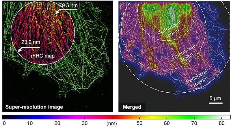
In a study published in Light: Science & Applications, a team of scientists have introduced a novel method known as the rolling Fourier ring correlation (rFRC). This method facilitates the representation of resolution heterogeneity directly in the Super Resolution (SR) domain, thereby enabling mapping at an unparalleled SR scale and an effortless correlation of the resolution map with the SR content.
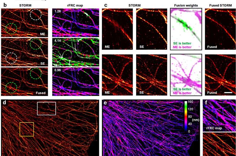
In addition, the team developed an improvement on the resolution scaled error map (RSM), resulting in more accurate systematic error estimation. This was used in tandem with the rFRC, creating a combined technique referred to as PANEL (Pixel-level Analysis of Error Locations), which focuses on pinpointing low-reliability regions from SR images.

The scientists successfully applied PANEL in a variety of imaging approaches, including Single-Molecule Localization Microscopy (SMLM), Super Resolution Radial Fluctuations (SRRF), Structured Illumination Microscopy (SIM), and deconvolution methods, verifying the effectiveness and stability of their quantitative map.
PANEL can be used to improve SR images. For instance, it has been effectively used to fuse SMLM images reconstructed by various algorithms, providing superior quality SR images.
In anticipation of their method becoming a staple tool for local quality evaluation, the team has made PANEL accessible as an open-source framework. Related libraries for MATLAB and Python are available, as well as a ready-to-use Fiji/ImageJ plugin on GitHub.
Further details about this promising technique can be found in a behind-the-scenes post written by the core team member Weisong Zhao, accessible here.
More information: Weisong Zhao et al, Quantitatively mapping local quality of super-resolution microscopy by rolling Fourier ring correlation, Light: Science & Applications (2023). DOI: 10.1038/s41377-023-01321-0
Journal information: Light: Science & Applications
Provided by Chinese Academy of Sciences


