This article has been reviewed according to Science X's editorial process and policies. Editors have highlighted the following attributes while ensuring the content's credibility:
fact-checked
peer-reviewed publication
trusted source
proofread
Single-particle photoacoustic vibrational spectroscopy using optical microresonators

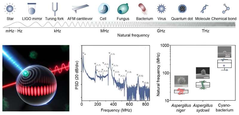
Pythagoras first discovered that the vibrations of strings are drastically enhanced at certain frequencies. This discovery forms the basis of our tone system. Such natural vibrations ubiquitously exist in objects regardless of their size scales and are widely utilized to derive their species, constituents, and morphology. For example, molecular vibrations at a terahertz rate have become the most common fingerprints for the identification of chemicals and the structural analysis of large biomolecules.
Recently, natural vibrations of particles at the mesoscopic scale have received growing interest, since this category includes a wide range of functional particles, as well as most biological cells and viruses. However, natural vibrations of these mesoscopic particles have remained hidden from existing technologies.
These particles with sizes ranging from 100 nm to 100 μm are expected to vibrate faintly at megahertz to gigahertz rates. This frequency regime could not be resolved by current Raman and Brillouin spectroscopies, however, due to strong Rayleigh-wing scattering, while the performances of piezoelectric techniques that are widely exploited in macroscopic systems degrade significantly at frequencies beyond a few megahertz.
In a new paper published in Nature Photonics titled "Single-particle photoacoustic vibrational spectroscopy using optical microresonators," a team led by Professor Xiao Yunfeng at Peking University has demonstrated the real-time measurement of natural vibrations of single mesoscopic particles using optical microresonators, extending the reach of vibrational spectroscopy to a new spectral window.
The working principle of microresonator-based vibrational spectroscopy is summarized according to Dr. Tang Shuijing as follows: "Mesoscopic particles are expected to vibrate at MHz to GHz rates, and their vibrational amplitudes are usually too subtle to be detected by traditional techniques. To address this issue, a new vibrational spectroscopy has been proposed. It involves using a short laser pulse to heat the particle and induce its vibrations.
"By directly placing the particle onto a high-Q optical microresonator, the vibrations of the particle generate acoustic waves within the microresonator, ultimately perturbating its optical mode," says Shuijing, a research associate professor at Peking University.
During the vibrational spectroscopy experiments, the researchers deposited mesoscopic particles on a silica microspherical resonator with a radius of approximately 30 μm and a quality factor of around 106. They then used a pulsed laser (with a wavelength of 532 nm and a duration of 200 ps) to irradiate particles and stimulate their vibrations, with the incident energy density of approximately 2 pJ μm−2.
A continuous-wave probe laser was coupled to the microresonator to excite its optical mode, and the vibrations of the particles were detected in real-time by monitoring the power of the transmitted laser. By using Fourier transformation of the temporal responses, the vibrational spectra of particles were obtained.
The vibrational spectroscopy was successfully verified using mesoscopic particles with different constituents, sizes, and internal structures. The results showed an unprecedented signal-to-noise ratio of 50 dB and a detection bandwidth over 1 GHz.
With this innovative technology, the team further demonstrated biomechanical fingerprinting of the species and living states of microorganisms at the single-cell level. They found that the natural frequencies of microbial cells of the same species are bunched, forming unique fingerprints due to the highly defined and stable morphology of certain biological species.
"This vibrational spectroscopy enables the interrogation of structures and mechanical properties of particles in a non-destructive way. Specifically, the important biomechanical properties of cells, which are related to their species and living states, can be inferred from the vibrational spectra," said Dr. Xiao Yunfeng, who is a Boya Professor at Peking University.
"This technology allows vibrational spectroscopy of a wide range of mesoscopic particles, which could revolutionarily advance our understanding of the mesoscopic world with unprecedented precision," he added.
Living cells are complex biosystems, and their mechanical properties play an important role in cellular function, development and disease. This work provides a new fingerprinting technology to study biological systems at the single-cell level, and would lead to new insights and discoveries in various scientific fields.
More information: Shui-Jing Tang et al, Single-particle photoacoustic vibrational spectroscopy using optical microresonators, Nature Photonics (2023). DOI: 10.1038/s41566-023-01264-3
Journal information: Nature Photonics
Provided by Peking University


