This article has been reviewed according to Science X's editorial process and policies. Editors have highlighted the following attributes while ensuring the content's credibility:
fact-checked
peer-reviewed publication
trusted source
proofread
Low-cost smartphone fluorescence microscope developed
 Illustration shows the light path for transmitted light viewing. Components shown include the smartphone, clip-on lens (black), glowscope frame (gray), stage and viewing platform (transparent), petri dish containing a specimen, and a LED work lamp positioned under the stage and viewing platform. (b) Image view using a smartphone camera without the clip-on lens shows zebrafish embryos (blue arrow, 3 dpf). A standard pencil (eraser side) is shown as a size reference. (c) Image view using the additional clip-on lens shows the same zebrafish embryos and pencil eraser seen in panel (b). Images shown in panels (b–c) are native magnification (not stretched) as seen on live-view, acquired using an Apple iPhone 12 Pro with 1x (middle magnification) lens, 6 × digital zoom. Credit: Schaefer et al., Scientific Reports (2023).")
A device that can convert a smartphone or tablet into a fluorescence microscope for less than US $50 is presented in a proof-of-principle study in Scientific Reports. The authors suggest that the device—which they have named a glowscope—could be used to image cells, tissues, and organisms under low magnification in schools, science outreach settings, and some research labs.
Fluorescence microscopes are used to study specimens labeled with fluorescent stains or expressing fluorescent proteins, such as those tagged with green fluorescent protein. However, as these microscopes usually cost at least several thousand US dollars, their use is typically limited to well-funded research labs.
The glowscope, devised by Jacob Hines and colleagues, is made of a plexiglass and plywood frame, a clip-on camera lens, an LED torch, and theater stage lighting filters. The frame is used to position a smartphone or tablet above a specimen and the lens is clipped onto the phone or tablet camera to enable magnification. The specimen is illuminated by the LED torch and a lighting filter is placed over the lens to filter out unwanted wavelengths of light and allow visualization of fluorescent light emitted by the specimen.
The authors demonstrated the abilities of the glowscope by using it to image live zebrafish embryos—which are between two and three millimeters long—expressing fluorescent proteins in either the spinal cord, cardiac tissue, or hindbrain. They found that the clip-on lens provided approximately five-fold magnification and was capable of imaging green and red fluorescent tissues with up to ten micrometer resolution—sufficient to view individual pigment cells. The authors used the glowscope to measure the heart rates of embryos and, after enhancing the clarity of videos recorded using free software, the movements of individual heart chambers.
-
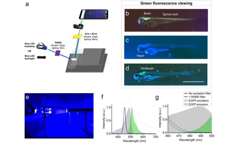
Use of recreational LED flashlights and theater stage lighting filters for smartphone green fluorescence microscopy. (a) Schematic of components used for green fluorescence viewing on the glowscope. (b–d) Representative fluorescence images of transgenic zebrafish embryos (3–4 dpf, lateral views) expressing green fluorescence in cell-type specific patterns. Reporter lines viewed include Tg(nkx2.2a:memGFP) (b), Tg(myl7:EGFP) (c), and Tg(phox2b:EGFP) (d). (e) Image shows the glowscope in use for green fluorescence viewing. (f–g) Plots show the blue LED flashlight emission wavelength (black and blue lines, measured using a Vernier spectrometer) in comparison to the excitation profile of EGFP (gray, obtained from www. fpbase. org). The dashed box region in panel (f) is further magnified in panel g to show the effect of the R4990 filter with greater detail. Images in panels (b–d) were acquired using an Apple iPhone XR. Credit: Schaefer et al., Scientific Reports (2023). -
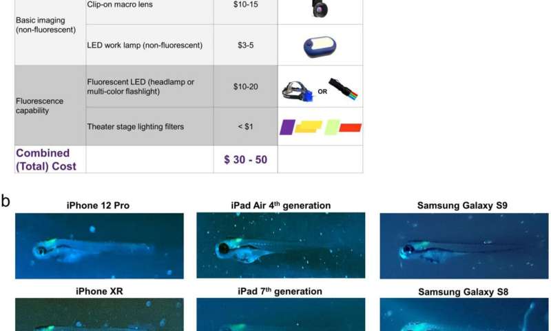
Summary of glowscope components and smartphone compatibility. (a) Table shows the basic components and costs (at the time of this study) in US Dollars. (b) Representative images of Tg(phox2b:EGFP) zebrafish embryos demonstrate compatibility with all devices tested, but subtle differences between various tablets and smartphones used for testing. Scale bar is 1 mm. Credit: Schaefer et al., Scientific Reports (2023).
As the materials for a glowscope cost between US $30 and $50, the authors propose that they could be used by school students to study anatomy, behavior, physiology, development, and genetic inheritance in small organisms expressing fluorescent proteins, which could be obtained from laboratories. Several glowscopes and smartphones could also be used simultaneously to acquire video data in research labs that do not have access to multiple fluorescence microscopes, they add.
A complete description of the parts and instructions needed to assemble a glowscope is available in the supplementary information of the paper.
More information: Jacob Hines, A low-cost smartphone fluorescence microscope for research, life science education, and STEM outreach, Scientific Reports (2023). DOI: 10.1038/s41598-023-29182-y. www.nature.com/articles/s41598-023-29182-y
Journal information: Scientific Reports
Provided by Nature Publishing Group


