Researchers realize single full field-of-view reconstruction fourier ptychographic microscopy

Fourier ptychographic microscopy (FPM) is a recently developed computational imaging technique, which has high-resolution and wide field-of-view (FOV). However, due to the lower light efficiency of the off-axis LEDs, the exposure time of dark-field images has to be extended to improve the signal-to-noise of dark-field images. In addition, effected by the spherical illumination wavefronts of LEDs, the wavevectors of full-FOV are different.
Therefore, the full-FOV has to be divided into sub-fields and reconstructed sequentially, and then stitch them to obtain a full-FOV high-resolution images. It is necessary to develop a new illumination method to provide plane wave illumination with uniform intensity and different angles.
In a study published in Biomedical Optics Express, a research group led by Prof. MU Quanquan from the Changchun Institute of Optics, Fine Mechanics and Physics of the Chinese Academy of Sciences realized a single full-FOV reconstruction FPM, which is termed full-FOV Fourier ptychographic microscopy (F3PM).
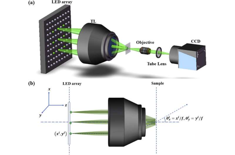
This novel illumination method is achieved by combing LED array and telecentric lens.
The role of telecentric lens is to collect the wavefronts from LEDs and collimates them into plane waves. The telecentric character and excellent plane wavefront of telecentric lens are the key elements in wavefront modulation. Excellent plane wavefront guarantees that the wavevectors are the same for full-FOV and the reconstruction process becomes more flexible, therefore the reconstruction size can be larger, and even the single full-FOV reconstruction can be implemented.
For conventional FPM, the full-FOV images reconstruct process consists of multiple reconstructions, intensity correction for different sub-fields and image stitching. In order to meet the needs of image stitching and light intensity correction, the overlap rate between adjacent sub-fields should be guaranteed 30% or more.
Compared with the conventional FPM, F3PM improves the size of single reconstruction from 0.25μm2 to 14.6 mm2, and eliminates the steps of image stitching and calculation redundancy. Without these steps, the reconstruction process for full-FOV high-resolution images becomes simpler. Based on multi-coding light scheme and wavefront modulation of telecentric lens, the single full-FOV reconstruction enables the dynamic imaging of FPM.
More information: Youqiang Zhu et al. Single full-FOV reconstruction Fourier ptychographic microscopy, Biomedical Optics Express (2020). DOI: 10.1364/BOE.409952
Journal information: Biomedical Optics Express
Provided by Chinese Academy of Sciences


