New contrast agent combination could usher in a new era of vascular systems biology

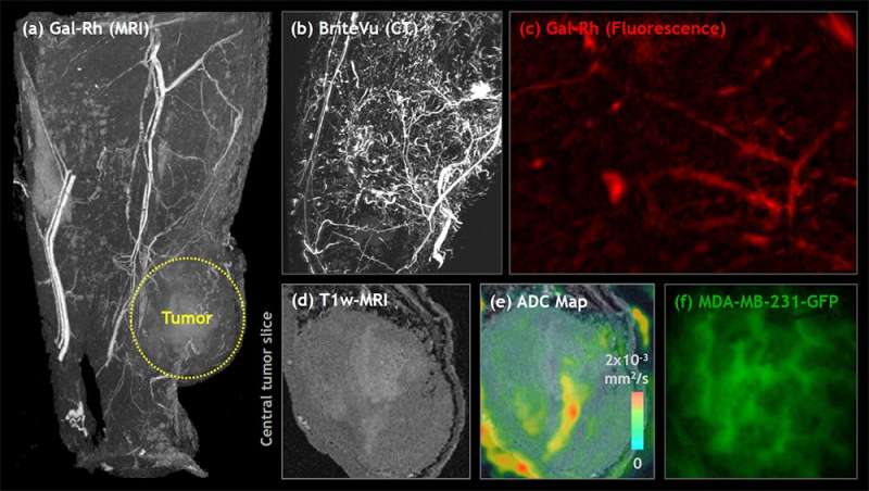
A new contrast agent combination for imaging tumor samples enhances imaging and data extraction and thus benefits image-based modeling of tumor processes. The discovery will be presented today at the American Physiological Society (APS) Interface of Mathematical Models and Experimental Biology: Role of the Microvasculature Conference in Scottsdale, Ariz.
Image-based systems biology combines images of the subject of study with computer and mathematical modeling to gain a fuller understanding of the underlying biological processes at multiple levels. For example, in vascular systems biology, researchers could extract data about the diameters, shapes and paths of connecting blood vessels to calculate factors such as the movement of fluid and cells through those vessels. When looking at the vasculature around and inside a tumor, such simulations could reveal important information about how the tumor extracts nutrients from the body, how it metastasizes or how to better deliver drugs and target therapies.
One difficulty with imaging tumors has been that employing a contrast agent necessary for one imaging method could preclude the use of another. Akanksha Bhargava, Ph.D., works in the lab of Arvind Pathak, Ph.D., at Johns Hopkins University (www.pathaklab.org) in Baltimore. Pathak has made strides in combining data from multiple imaging techniques and spatial scales to develop a more complete representation of the tumor environment. Bhargava explains that "this new combination of a radio-opaque CT agent, … magnetic resonance-visible and fluorescent agents does not interfere with other image contrast mechanisms."
The team has already used the new combination of agents to assemble MRI, CT and optical imaging data of different 3-D tissues, allowing for visualization from the cellular- to whole-tissue level with complementary contrast mechanisms. They hope to use it to develop an "atlas for cancer systems biology" that curates digitized results of the images this approach will make possible. When completed, the contents of the digitized atlas would be free and downloadable for researchers around the globe.
"We believe this new approach will encourage novel applications and usher in a new era of image-based systems biology research," Pathak says.
More information: The APS Interface of Mathematical Models and Experimental Biology: Role of the Microvasculature will be held September 11–14 in Scottsdale, Ariz.
Provided by American Physiological Society


