March 31, 2016 report
Dipole-dipole interactions imaged at sub-molecular resolution for first time

(Phys.org)—A team of researchers with the University of Science and Technology in China has for the first time, imaged dipole-dipole interactions using scanning tunneling microscopy. In their paper published in the journal Nature, the team describes how they captured the imagery and why they believe they may have also captured an instance of entanglement of chromophores. Guillaume Schull with Institutde Physique et Chimie des Matériaux de Strasbourg outlines the work done by the team in a News & Views article published in the same journal issue and explains in more detail the importance of better understanding the interactions of dipoles in biological processes.
Molecules with atoms that have unequal numbers of electrons are known as dipoles—different sides can be positively or negatively charged causing interactions with other molecules to come about. Interactions between dipoles is a major area of research because it is part of such important processes as photosynthesis. In plants, dipole interactions aide chromophore couplings—helping to transfer energy from sunlight to other molecules that later convert it to energy. How exactly this process works is still not fully understood, unfortunately, and for that reason, researchers have been trying to capture images of it as it occurs—but until now, have been unsuccessful because light based microscopes can only see images to a certain small size. In this new effort, the researchers used scanning tunneling microscopy to get the job done.
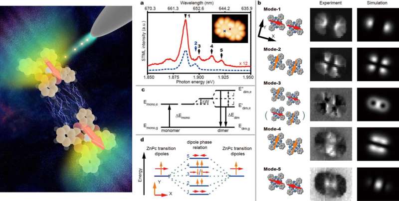
To capture the images, the researchers used chromophores that were made using a purple dye and then focused a red light on them to further highlight details. Next, they used the tip of the tunneling device to push some of the chromophores together—as they did so, they noticed that at approximately 3 nanometers apart, the light given off by the chromophores began to change; imagery of that change showed the process of dipole-dipole interactions taking place.
But, that was not the end of the story, in recent years some scientists have begun to suspect that entanglement occurs during dipole-dipole interactions. As part of their experiments, the researchers varied the numbers of chromophores involved in the coupling, trying clusters up to four in size. In so doing they found that the way that the light changed between them, might be an indication of entanglement.

More information: Yang Zhang et al. Visualizing coherent intermolecular dipole–dipole coupling in real space, Nature (2016). DOI: 10.1038/nature17428
Abstract
Many important energy-transfer and optical processes, in both biological and artificial systems, depend crucially on excitonic coupling that spans several chromophores. Such coupling can in principle be described in a straightforward manner by considering the coherent intermolecular dipole–dipole interactions involved. However, in practice, it is challenging to directly observe in real space the coherent dipole coupling and the related exciton delocalizations, owing to the diffraction limit in conventional optics. Here we demonstrate that the highly localized excitations that are produced by electrons tunnelling from the tip of a scanning tunnelling microscope, in conjunction with imaging of the resultant luminescence, can be used to map the spatial distribution of the excitonic coupling in well-defined arrangements of a few zinc-phthalocyanine molecules. The luminescence patterns obtained for excitons in a dimer, which are recorded for different energy states and found to resemble σ and π molecular orbitals, reveal the local optical response of the system and the dependence of the local optical response on the relative orientation and phase of the transition dipoles of the individual molecules in the dimer. We generate an in-line arrangement up to four zinc-phthalocyanine molecules, with a larger total transition dipole, and show that this results in enhanced 'single-molecule' superradiance from the oligomer upon site-selective excitation. These findings demonstrate that our experimental approach provides detailed spatial information about coherent dipole–dipole coupling in molecular systems, which should enable a greater understanding and rational engineering of light-harvesting structures and quantum light sources.
Journal information: Nature
© 2016 Phys.org


