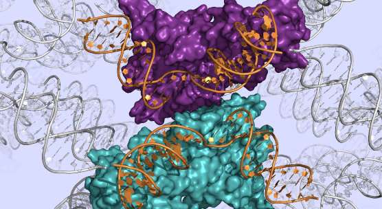
DNA repair enzyme mapped in atomic detail

An enzyme crucial to the process of DNA repair in our cells has been mapped in atomic detail by researchers at the University of Dundee, the UK's top-rated University for Biological Sciences.
DNA repair plays a key role in human diseases such as cancer. Researchers say that revealing the 3D molecular structure of a key enzyme involved in this process could be an important step towards developing future drugs.
DNA is the genetic `library' of the cell and is so important that it must be repaired when damaged. It is the only kind of molecule in the cell that is subject to such repair. In one such process, DNA temporarily forms interconnections, called junctions, that much be processed by special enzymes, including one called GEN1, which has now been mapped by the Dundee team.
"GEN1 was the first of these proteins to be identified, and only relatively recently," said Professor David Lilley, from the School of Life Sciences at the University of Dundee, who has led this new work.
"We have now determined the molecular structure of GEN1 in fine detail. This is an important step towards understanding DNA repair and how we may be able to develop drugs in future that can affect it."
To understand how an enzyme carries out its role in the cell, it is important to determine the structure of its molecules. This means working out how all its constituent atoms sit in 3D space.
This is achieved by obtaining small crystals of the protein, which then scatter X-rays generated by a huge particle accelerator, called a synchrotron. It is possible to reconstruct the 3D structure of the protein molecule from the resulting pattern of diffracted X-rays using mathematics.
Professor Lilley added, "In this study we used a form of the GEN1 enzyme from a kind of yeast that lives at high temperature. The enzyme is very closely related to the human protein, but has much better properties for study. This enabled us to study its biochemical properties in detail, and then go on to determine its structure by X-ray crystallography."
The resulting images of the molecular structure show GEN1 intimately bound to DNA, with extensive contacts between the two molecules.
"The whole shape of the protein has evolved to recognise the unusual shape of the DNA junction," said Professor Lilley.
This research is funded by Cancer Research UK, which has provided support to Professor Lilley's laboratory for over 20 years.
The research results have been published online in the journal Cell Reports.
Journal information: Cell Reports
Provided by University of Dundee


