A detector you can wear

Swiss scientists from ETH Zurich have developed the first elastic detector for magnetic resonance imaging (MRI). The detector in the form of an elastic bandage moulds itself to the shape of the patient's body, which also enables body parts to be examined in motion. The novel detector provides better images and greater patient comfort during the scan.
Ever since the early days of magnetic resonance imaging (MRI) around thirty years ago, all technical conditions have had to be as perfect and stable as possible for a successful scan: "You need a strong magnetic field that, until now, has had to be as homogeneous and constant as possible and the scans have to proceed exactly as planned," says Klaas Prüssmann, a professor at the joint Institute for Biomedical Engineering of ETH Zurich and the University of Zurich. "Nowadays, we want to shake off these constraints."
Not more powerful—more intelligent
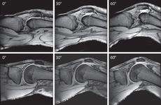
The aim is not only to keep making these devices more powerful, but also more intelligent. In their latest prototypes, Prüssmann says, the magnetic field is allowed to wobble thanks to measurement and control technology that handles even substantial field fluctuations. It should also be possible to examine body parts in motion, which is important to discern what the kneecap does while bending or whether the meniscus is torn partially or fully, for instance.
Present-day detectors might be robust and well-engineered, but they have two major shortfalls: they do not enable body parts to be scanned in motion and are built in such a way as to fit any patient. This means that the distance from the detector is vast for a small knee, which affects the detectors' sensitivity.
"It is as if a doctor didn't place his stethoscope directly on the body and tried to listen to the patient from ten centimetres away," says Prüssmann. For the first time, the ETH-Zurich professor and his team have now developed a detector that is both stretchable and elastic and can mould itself to the individual body.

An MRI scan of the knee, for instance, basically requires two things: a strong magnet and an antenna that emits radio waves and can serve as a detector. The latter surrounds the patient's knee while in the tube that builds up the magnetic field.
Detector like an elastic bandage
The detectors that record the signals are based on a resonant circuit. The largest module is a conductor loop with high conductivity. Making this both stretchable and elastic so that it returns to its original shape after stretching was a challenge for the scientists. Due to the high conductivity required, besides copper only gold and silver came into question, but they were too soft and not elastic enough.
Jurek Massner, one of Prüssmann's doctoral students, solved the problem as part of his dissertation. He sewed—as a conductor loop—fine copper braids onto a fabric blend of cotton and polyamide, which helped provide the necessary elasticity and stretchability. His detector resembles an elastic bandage.
The scientists had to make several prototypes until they found the ideal combination of conductor loop and support material. The electronic properties had to be distributed optimally in the conductor-loop braiding to provide the right resistance, inductance and capacitance there. In doing so, it became apparent that especially thin braiding had the best properties.
Eliminating interference with sophisticated electronics
The elastic detector's electronics presented a further challenge. When the detector is stretched out, the electrical conditions change and therefore so does the kind of signal. In order to be tolerant of such changes, Massner devised a special adjustment network and preamplifier to offset this "interference".
Until ten years ago, people mostly only worked with one detector in MRIs. Nowadays, as many receivers as possible are used in unison. Because every detector is in a slightly different position, they provide different perspectives of the same object. By exploiting these in the coding and signal processing, the imaging can be rendered considerably more rapid.
As a result, for instance, three-dimensional datasets with resolutions of up to 200 micrometres can be obtained. In the latest MRI machines with up to thirty-two detectors, the measuring time can be slashed to an eighth of what it used to be. Without speeding it up, such a scan would take over an hour.
Anyone who would like to find out more about the research conducted by Klaas Prüssmann and his team and even take a peek inside the MRI lab will have the opportunity to do so at Scientifica.
More information: Nordmeyer-Massner JA, De Zanche N, Pruessmann KP: Stretchable coil arrays: application to knee imaging under varying flexion angles. Magnetic Resonance in Medicine (2012), 67, 872-879, doi: 10.1002/mrm.23240
Provided by ETH Zurich


