New microscope may take the 'ouch' out of blood tests

If the sight of a needle makes you squeamish, researchers at the Technion-Israel Institute of Technology are developing a new optical microscope for viewing blood cells that could do away with conventional blood tests. The device, in an early prototype stage, would make it possible to collect vital blood information by simply shining a light through the skin to look directly at the blood.
The microscope’s benefits are manifold. Information is read immediately -- eliminating the waiting time for test results, and leading to earlier diagnoses. The microscope is widely accessible because it does not rely on medical labs to decipher the results. And crucial for the faint of heart – the 30-second procedure eliminates the use of needles.
Professor Dvir Yelin of the Technion Department of Biomedical Engineering and the Lorry Lokey Interdisciplinary Center for Life Sciences and Engineering heads the research team, whose work was published in the May 2012 issue of Biomedical Optics Express.
“We have invented a new optical microscope that can see individual blood cells as they flow inside our body,” says Lior Golan, a graduate student on the team.
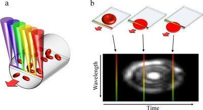
The device relies on a technique called spectrally encoded confocal microscopy (SECM), which allows for 2-dimensional spatial imaging of blood cells. It works by pressing a probe that generates a red to violet line spectrum of light against the patient’s skin. As moving blood cells that are near the surface of the skin cross the beam, they scatter the light’s rays, which are then collected by the probe and analyzed to generate 2D images of the blood cells.
Other blood-scanning systems with cellular resolution exist, but often rely on potentially harmful fluorescent dyes that must be injected into the patient’s bloodstream.
The device is still years away from clinical application, but the team is already working on a second-generation system that will be able to beam deeper into the body. Prof. Yelin’s prototype is the size of a shoebox, but the team hopes to have developed a thumb-sized prototype within a year.
Provided by American Technion Society


