This article has been reviewed according to Science X's editorial process and policies. Editors have highlighted the following attributes while ensuring the content's credibility:
fact-checked
proofread
Wide-field, high-resolution and broadband mesoscopic objective lens
 Structure of the mesoscopic objective, (b) Aberration distribution of the mesoscopic objective, (c) Wide-field imaging system built with the mesoscopic objective, (d) Laser point-scanning imaging system built with the mesoscopic objective. Credit: Opto-Electronic Advances (2024). DOI: 10.29026/oea.2024.230212")
Optical microscopes are indispensable research tools in fields such as life sciences, medical science, and materials science. The objective lens is the core component of the microscope, determining two key parameters of microscopic imaging: resolution and imaging field of view (FOV).
These two parameters are mutually constrained. Commercial microscope objectives with a numerical aperture (NA) of 0.5 offer submicron resolution; however, their imaging FOV is often limited to around 1 mm. The 2014 Nobel Prize in Chemistry was awarded for super-resolution microscopy, a technology that greatly enhances imaging resolution.
However, achieving high resolution and a large FOV simultaneously remains a research challenge. In recent years, the demand for cross-scale high-throughput imaging has been increasing, but conventional microscope objectives cannot simultaneously achieve both a large FOV and high resolution.
This makes high-resolution imaging of large samples difficult. The usual method is to image the sample multiple times in small FOV and then stitch the images together to form the desired imaging field. However, this method produces artifacts at the stitching edges and has a slow imaging speed, making it impossible to observe dynamic changes of the sample in real-time.
To address these issues, mesoscopic objectives have been proposed. They have complex optical structures and excellent aberration optimization, enabling high NA and a super-large imaging FOV, significantly enhancing the imaging throughput of optical microscopes.
In 2016, the University of Strathclyde first developed a mesoscopic objective lens with a 0.47 NA, a 6 mm FOV, and a working wavelength range from 400 nm to 700 nm. That same year, Physics World magazine selected it as one of the top ten physics breakthroughs of the year. Subsequently, related research has been reported internationally.
However, optimizing chromatic aberration over a large FOV is extremely challenging. Current mesoscopic objectives are limited to a single imaging wavelength band, either visible or near-infrared, and cannot meet the requirements for diverse fluorescence imaging, such as single-photon or two-photon imaging. Additionally, the FOV of existing mesoscopic objectives is concentrated in the range of 3 mm to 6 mm. Increasingly, application scenarios demand further enhancement of the imaging FOV to achieve higher imaging throughput.
To overcome the current obstacles of narrow imaging wavelength band and insufficient imaging FOV in mesoscopic objectives, a research group led by Prof. Guohua Shi from the Suzhou Institute of Biomedical Engineering and Technology, Chinese Academy of Sciences, designed a flat-field apochromatic objective structure for mesoscopic fields and developed the world's largest imaging field and broadest working wavelength mesoscopic objective with submicron resolution.
It features an 8 mm diameter FOV, 0.5 NA, and an imaging wavelength range of 400-1000 nm, with the unique ability to image in both visible and near-infrared wavelengths. The effort is reported in Opto-Electronic Advances.
-
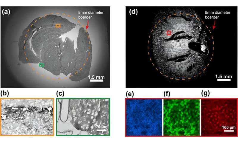
(a-c) Imaging results of mouse brain, (d-g) Fluorescence imaging results of mouse kidney. Credit: Opto-Electronic Advances (2024). DOI: 10.29026/oea.2024.230212 -
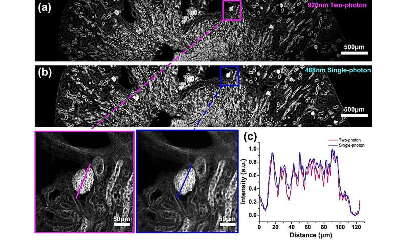
(a) Point-scanning two-photon fluorescence imaging results, (b) Point-scanning single-photon fluorescence imaging results, (c) The intensity distribution curves of the same glomerular in (a) and (b). Credit: Opto-Electronic Advances (2024). DOI: 10.29026/oea.2024.230212
The objective consists of 10 groups of 19 spherical lenses, using a three-group aberration optimization design.
The front group includes a doublet lens and a triplet lens with an approximate aplanatic design, achieving the required NA while minimizing aberrations. The middle group comprises a single lens, a doublet lens, a triplet lens, and two doublet meniscus lenses in a double Gauss configuration, primarily correcting the aberrations of the front group. The rear group includes two single lenses and a doublet lens for correcting residual aberrations.
For chromatic aberration optimization, the front group uses low-dispersion glass to reduce chromatic aberration. The middle group's symmetric meniscus lens structure compensates for lateral chromatic aberration, and the triplet lens compensates for axial chromatic aberration.
The rear group employs a doublet structure with similar refractive indices but significantly different dispersions, and large air gaps to compensate for chromatic aberration. The combined structural designs meet the aberration optimization requirements, ultimately achieving apochromatic correction from visible to near-infrared wavelengths in mesoscopic fields.
To verify the imaging performance of the objective, the research team conducted various performance tests. Imaging the resolution test chart with this objective measured a bright-field resolution of 714 lp/mm and a fluorescence microsphere resolution of 0.74 μm, with a field distortion of 0.46%.
When imaging mouse brain and kidney slices, a 1.35-billion-pixel per frame image was obtained. A quantitative comparison with a commercial 20 x 0.5 NA objective showed that the mesoscopic objective has similar image quality but over 40 times the imaging field area of the commercial objective.
To verify the scanning imaging capability of the objective, the team built a laser point-scanning mesoscopic imaging system, obtaining single-photon fluorescence imaging results with 488 nm continuous light excitation and two-photon fluorescence imaging results with 920 nm femtosecond pulse light excitation.
Single-photon imaging provides higher signal intensity, while two-photon imaging offers higher imaging contrast. This is also the first time that simultaneous single- and two-photon imaging has been achieved with a mesoscopic objective.
This objective has significant potential for high-resolution imaging of large-scale samples, such as brain mapping, cross-brain region single- and two-photon imaging, and high-resolution imaging of organoids.
More information: Xin Xu et al, Large-field objective lens for multi-wavelength microscopy at mesoscale and submicron resolution, Opto-Electronic Advances (2024). DOI: 10.29026/oea.2024.230212
Provided by Compuscript Ltd


